goals and approach
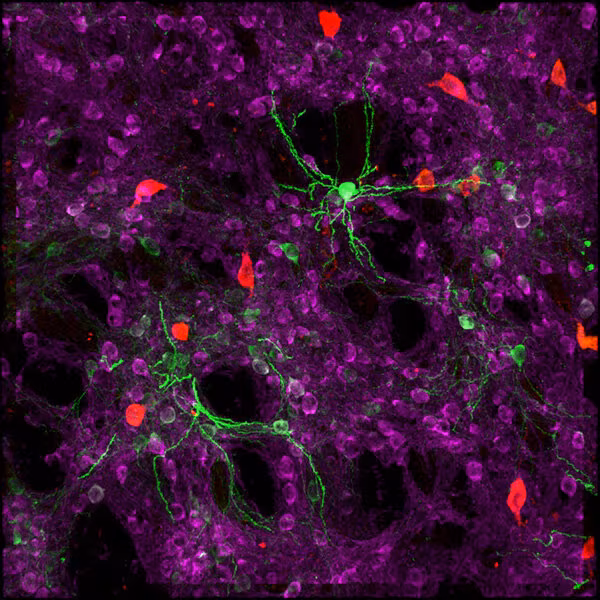
The Imaging team at the Allen Institute's brain science accelerator is revealing the organization of cell types in mammalian brains in aging and disease. This team uses many imaging techniques including confocal microscopy, brightfield microscopy, 2- and 3-photon imaging, spatial transcriptomics, and lightsheet microscopy in cleared tissue.
The Imaging team is grouped into sub-teams, each focused on different technologies. The spatial transcriptomics team images mRNA molecules to identify and locate cell types in tissue sections. The scanning lab team characterizes patterns of gene expression, cell types, and microcircuits using high-throughput brightfield and fluorescence slide-scanning microscopes. The whole brain imaging team develops and operates 2-photon and lightsheet microscopes that image fluorescence throughout intact mouse brains. The three-photon team is improving optical methods for deep cellular calcium imaging in mouse and non-human primate brains in vivo.