lessons & resources in this unit
in this lesson





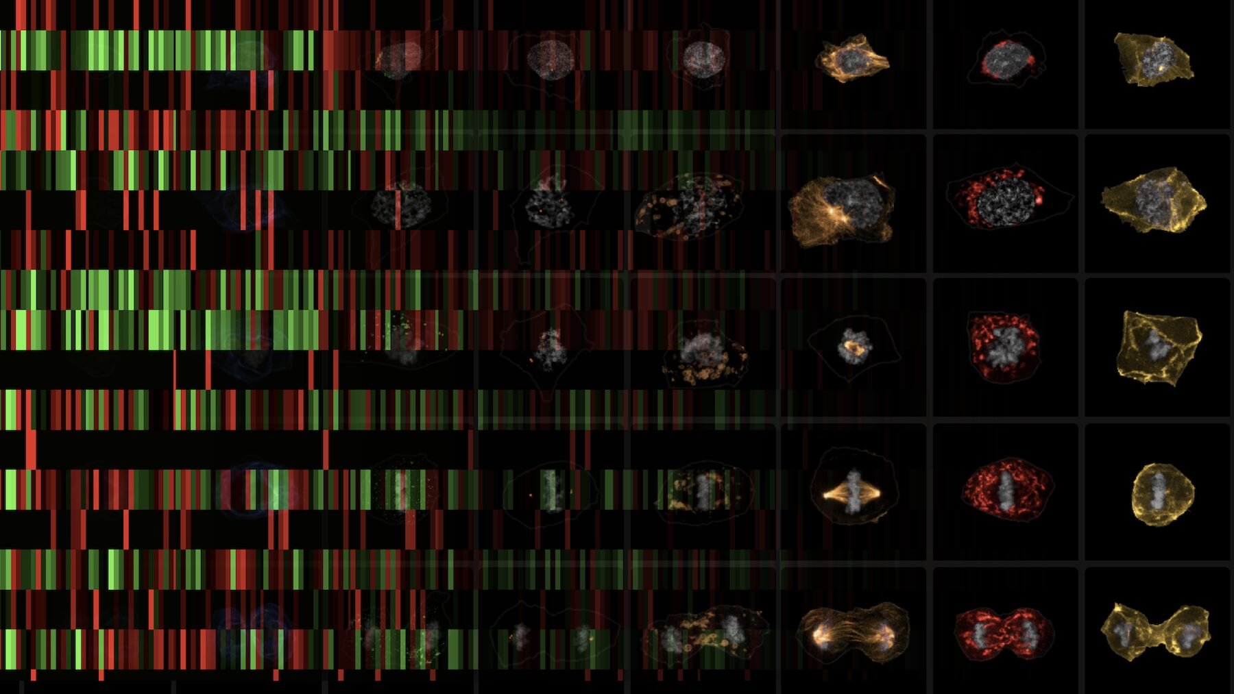
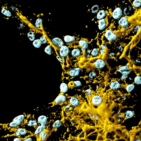
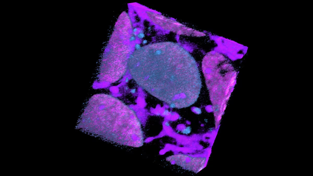
This lesson introduces students to research at the Allen Institute involving fluorescently tagged human induced pluripotent stem cells (hiPSCs). Students then learn how to use a web-based tool, the Cell Feature Explorer (CFE), which allows them to explore open access images and data from hundreds of thousands of fluorescently labeled hiPSCs throughout the cell cycle.
learning objectives/
- Define human induced pluripotent stem cells (hiPSCs)
- Describe how fluorescent microscopy images are collected and processed at the Allen Institute for Cell Science
- Observe images of cells undergoing mitosis using the Cell Feature Explorer
- Interpret cell images to classify them by cell cycle stage
Take a look inside our labs as Allen Institute scientists walk through the process of developing their fluorescently tagged human induced pluripotent stem cell lines, growing and maintaining those cells, and taking images for their research
materials
- Instructor Guide
- Slides
- Worksheet & KEY
additional resources/
terms of use/
This work is licensed under the Creative Commons Attribution-NonCommercial-ShareAlike 4.0 International (CC BY-NC-SA 4.0). To view a copy of this license, visit https://creativecommons.org/licenses/by-nc-sa/4.0/.
The development of this lesson was supported by the National Science Foundation under Award Number 2236366. The content is solely the responsibility of the authors and does not necessarily represent the official views of the National Science Foundation.
Last updated March 2026