lessons & resources in this unit
in this lesson





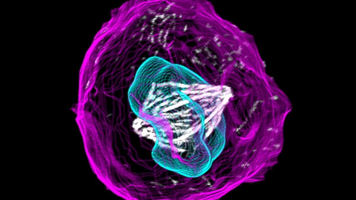
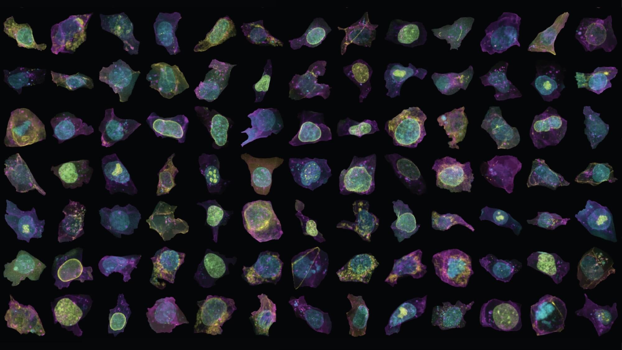
This lesson introduces students to research at the Allen Institute exploring intracellular organization within fluorescently tagged human-induced pluripotent stem cells (hiPSCs). After learning about fluorescently tagged hiPSCs, students are introduced to the Deep Cell Zoom online tool where they can view over 200,000 images of these cells. Students are then asked to explore the collection of images and visually identify examples of cells at each stage of mitosis. At the end of this lesson, students will have created their own cell gallery featuring real cell images representing the different stages of mitosis.
learning objectives/
- Define human induced pluripotent stem cells (hiPSCs)
- Describe how fluorescent microscopy images are collected and processed at the Allen Institute
- Explore real images of human induced pluripotent stem cells from the Deep Cell Zoom Tool
- Categorize cell images based on what stage of the cell cycle/mitosis they appear to be in
- Justify the images you chose for your cell gallery by articulating what visual aspects of a cell made you classify it as a certain stage of mitosis
terms of use/
This work is licensed under the Creative Commons Attribution-NonCommercial-ShareAlike 4.0 International (CC BY-NC-SA 4.0). To view a copy of this license, visit https://creativecommons.org/licenses/by-nc-sa/4.0/.
funding acknowledgement/
The development of this lesson was supported by the National Science Foundation under Award Number 2236366. The content is solely the responsibility of the authors and does not necessarily represent the official views of the National Science Foundation.
Last updated March 2026