lessons & resources in this unit
in this lesson





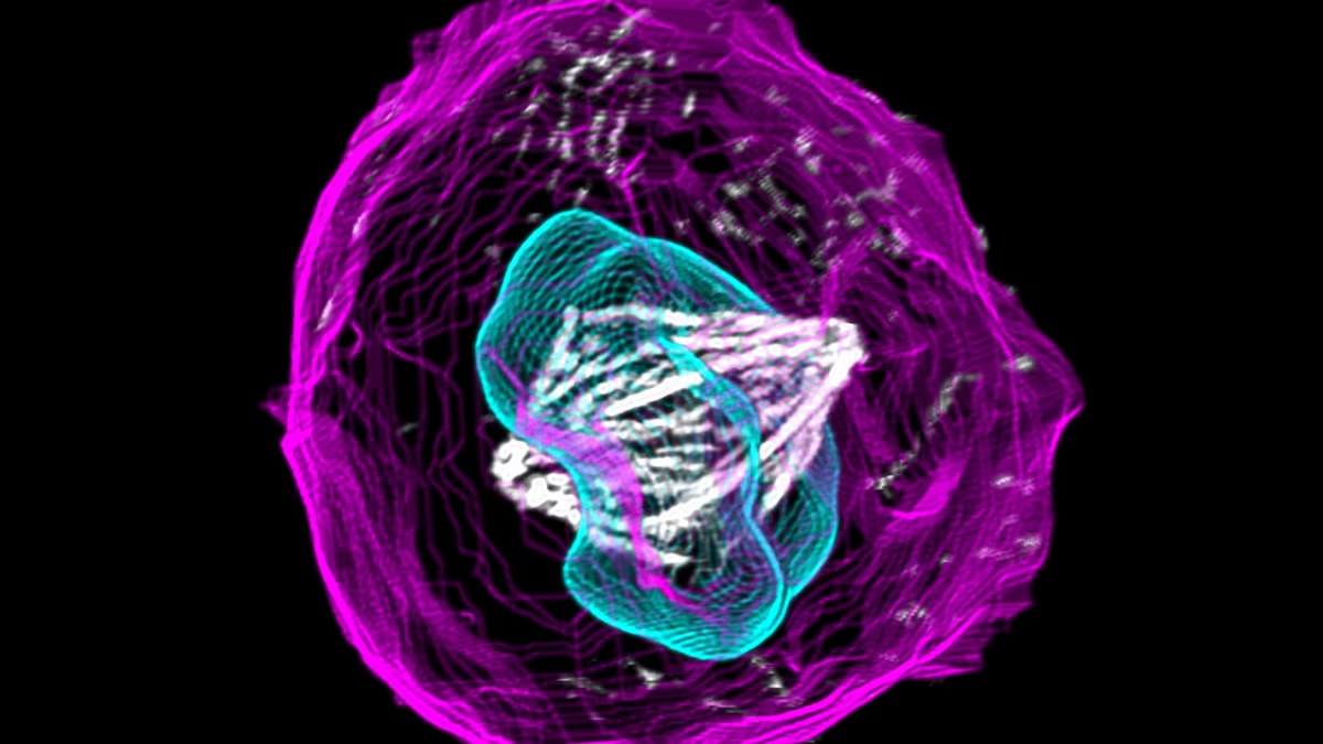
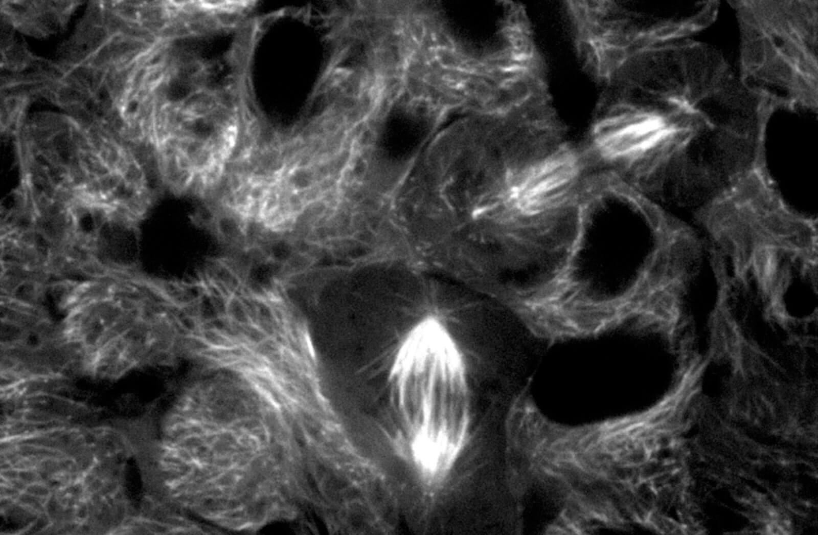
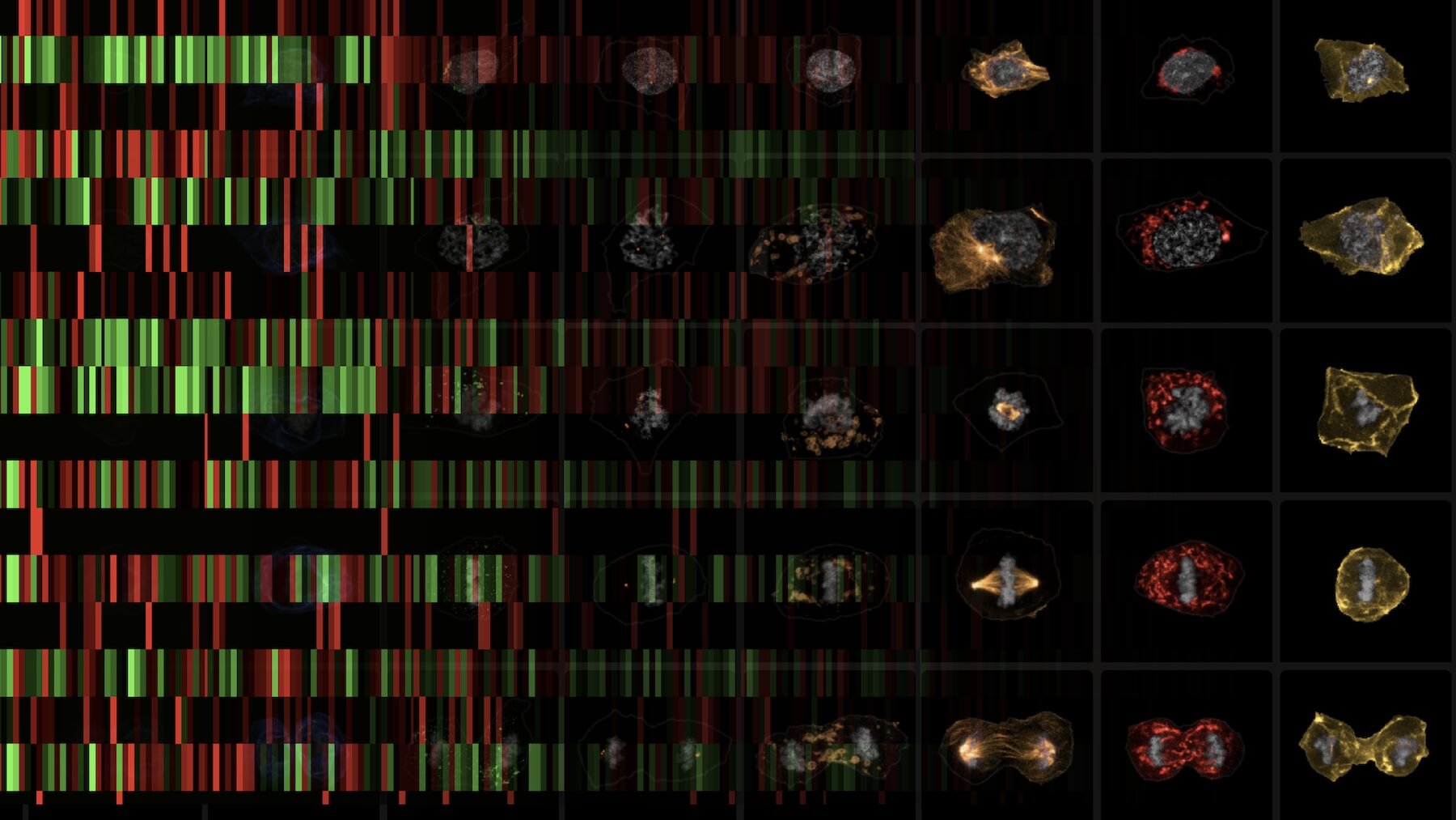
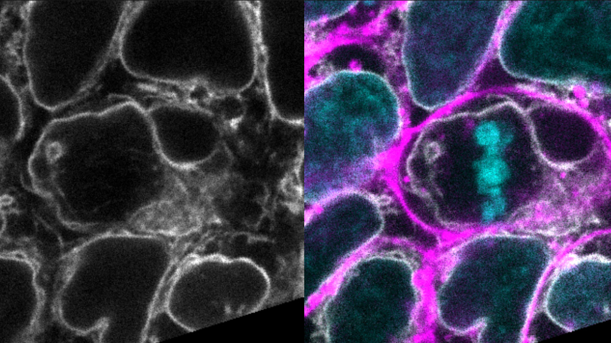
This lesson challenges students to navigate the Integrated Mitotic Stem Cell, an interactive, open access tool that contains image data from a spinning disc confocal microscope of human induced pluripotent stem cells with different structures fluorescently tagged. The lesson includes an accompanying worksheet that guides students through the tool and prompts them to explore cell structure localization by analyzing z-stacks of real cell images from different stages of mitosis.
What is a Z-stack?
A Z-stack is a composite image where multiple images of the same object at different depths are taken sequentially under a microscope. These images can then be stitched together to allow you to explore the imaged object in three dimensions.
learning objectives/
- Explore elements of mitosis by looking at structures beyond DNA and microtubules
- Review hiPSCs, fluorescent tagging, and cell colonies
- Synthesize information about organelle localization from Z-stack images
- Analyze the relative abundance of different cell structures within various parts of the cell (top, middle, bottom)
terms of use/
This work is licensed under the Creative Commons Attribution-NonCommercial-ShareAlike 4.0 International (CC BY-NC-SA 4.0). To view a copy of this license, visit https://creativecommons.org/licenses/by-nc-sa/4.0/.
The development of this lesson was supported by the National Science Foundation under Award Number 2236366. The content is solely the responsibility of the authors and does not necessarily represent the official views of the National Science Foundation.
Last updated March 2026