in this article
With the sun setting earlier in the winter months, you may be walking your dog or taking a post-work jog in the dark. On one of these neighborhood strolls, you may round a corner and be startled to see another nighttime walker in your path. Without thinking about it, your brain will cue you into that faint presence so you can quickly move a respectful 6 feet out of their way. A new study has pinpointed the neurons that may underlie the brain’s response to weak but relevant visual cues, like a figure in the dark.
Researchers at the Allen Institute recently published a study in the journal eLife that outlined the role of these neurons, known as VIP interneurons, in the mouse visual system. They found that VIP interneurons seem to be specifically tuned to subtle — but likely still important — visual cues. Given that these interneurons are well conserved across mammalian species, there’s good reason to suspect that they kick into gear when we encounter a faint obstacle as well.
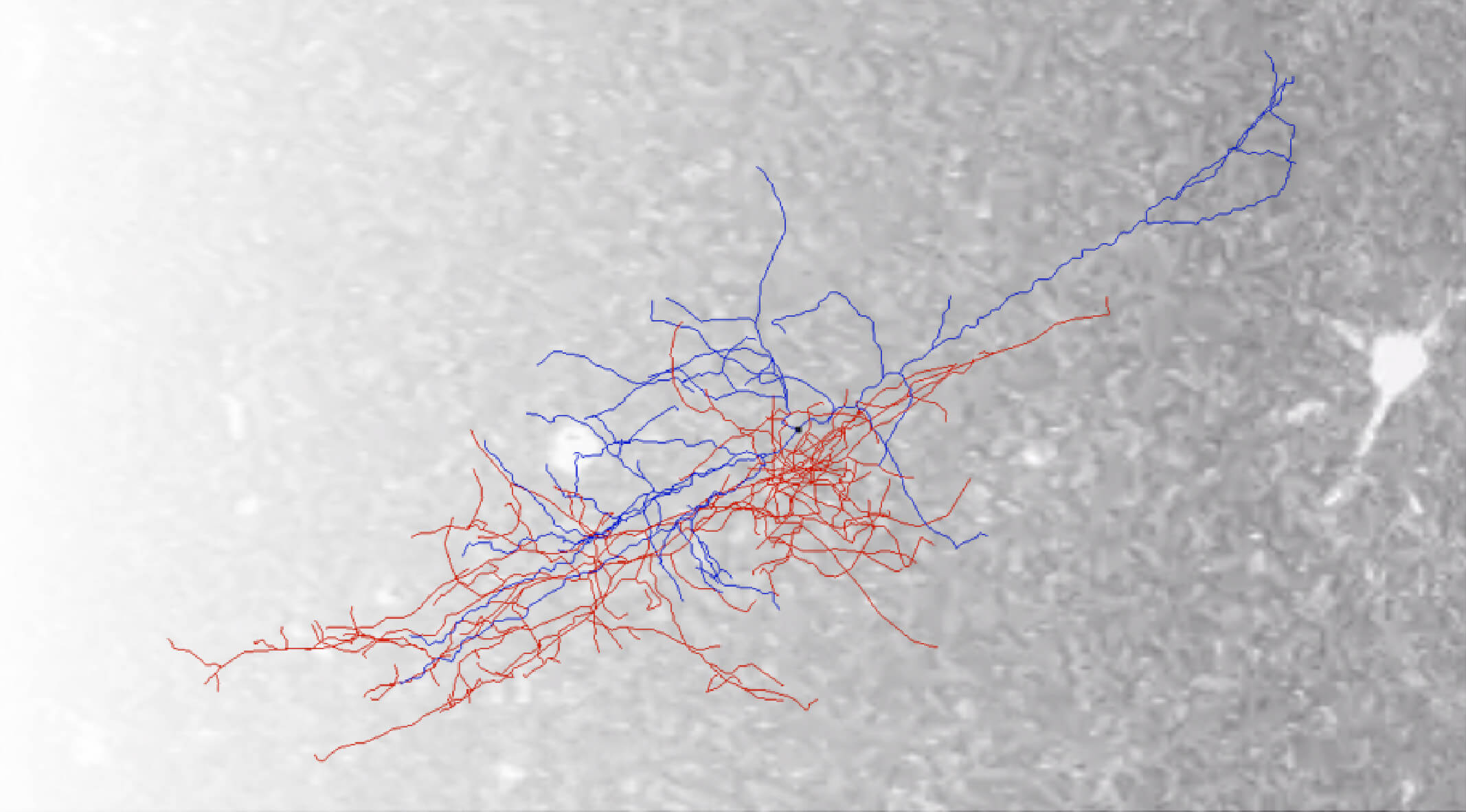
To examine their function, Allen Institute researchers focused on the cortex, the outermost shell of the brain concerned with things like attention, thought, and consciousness. For decades, scientists have known that different neurons in the visual cortex, the region that responds to what we see, are tuned to specific features in our field of view. For example, certain neurons respond strongly when encountering a horizontal or a vertical line or edge. Other neurons can be tuned to switch on in response to size, with some neurons responding to small objects while others respond to large objects. When a neuron recognizes a visual cue that it is tuned to, it fires a rapid electrical response. At the Allen Institute, researchers have measured this neuronal activity in the mouse visual system to determine the different kinds of visual cues, also known as stimuli, that evoke responses in an experimental survey known as the Allen Brain Observatory.
The mice in the Allen Brain Observatory are genetically engineered so specific neurons glow under a fluorescent microscope when the cells are electrically active. For the eLife study, mice were walking or running at will on a platform, like a tiny treadmill, and presented with images of greyscale bars that were either bold (“high contrast”) or faint (“low contrast”). Researchers were able to better understand how the mouse brain perceives its environment by seeing which neurons glowed in response to certain bars, and if that activity changed when the mice were running.

“We want to understand how the visual cortex responds given some input from the outside world, but we also want to understand how that response is going to be changed by the behavioral state,” said Dan Millman, Ph.D., a Scientist in the Allen Institute MindScope Program and first author on the study. “Both aspects are very important.”
Millman and his colleagues found that, unlike most neurons in the mouse visual cortex, VIP interneurons were actively suppressed by other neurons whenever a bold stimulus was present in the mouse’s field of view. VIP interneurons were only activated when they were shown a faint stimulus. Their activity increased even more in response to the low contrast stimulus when the mouse was running.
“If you’re a mouse, running, and something is approaching head on, you want to be sensitive to that,” said Millman, who led the study along with Allen Institute Assistant Investigator Saskia de Vries, Ph.D. “You don’t want to miss a predator or an obstacle.”
You can imagine yourself in a similar scenario; if you’re running at night, it becomes more important to spot obstacles in your path. When the contrast, or boldness, of a visual cue is high, detecting it is no problem. But when that contrast is low, the brain needs a different sensory strategy.
VIP interneurons are a type of inhibitory neuron, a class of neurons that suppress other neurons’ activity. Previously, Allen Institute researchers hypothesized that VIP interneurons help the brain recognize novel images. They quiet other inhibitory neurons, releasing the brakes on excitatory neurons to elevate their activity under certain circumstances. In their current study, the researchers found that when VIP interneurons are active, they also boost the overall response of excitatory neurons to the weak stimuli. In the case of a faint obstacle, VIP interneurons might thus free excitatory neurons to notice and react to a possible threat.
The Allen Institute researchers believe VIP interneurons could play a similar role in detecting other kinds of weak signals, such as a quiet whisper or a faint smell.
“We think that the circuit is canonical and repeats throughout the cortex. If we can understand how VIP interneurons work in the visual cortex, we’re hoping that can lead us to more general insights for the whole cortex,” said Millman.
Just as they are activated by faint images in the visual cortex, researchers have shown that VIP interneurons are activated by faint noises in the auditory cortex and hypothesize that they respond to weak transmissions in other brain regions. By recognizing weak signals and directing the brain’s attention to them, VIP interneurons could be catching what we might otherwise miss, everything from a tripping hazard in the dark living room to the patter of your child’s feet during a Zoom call. — written by Anna Marie Yanny
Citations
about the allen institute
The Allen Institute is an independent, 501(c)(3) nonprofit research organization founded by philanthropist and visionary, the late Paul G. Allen. The Allen Institute is dedicated to answering some of the biggest questions in bioscience and accelerating research worldwide. The Institute is a recognized leader in large-scale research with a commitment to an open science model. For more information, visit alleninstitute.org.


