discover
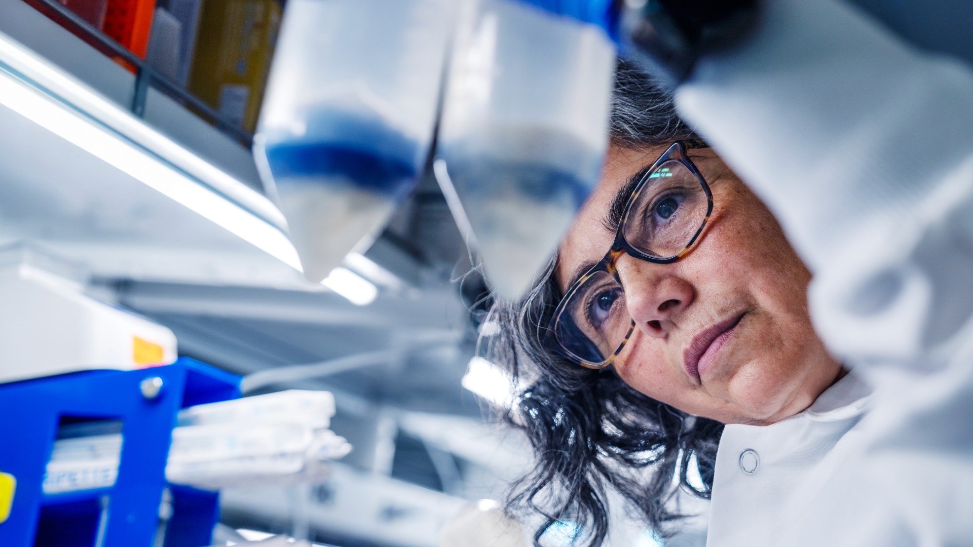
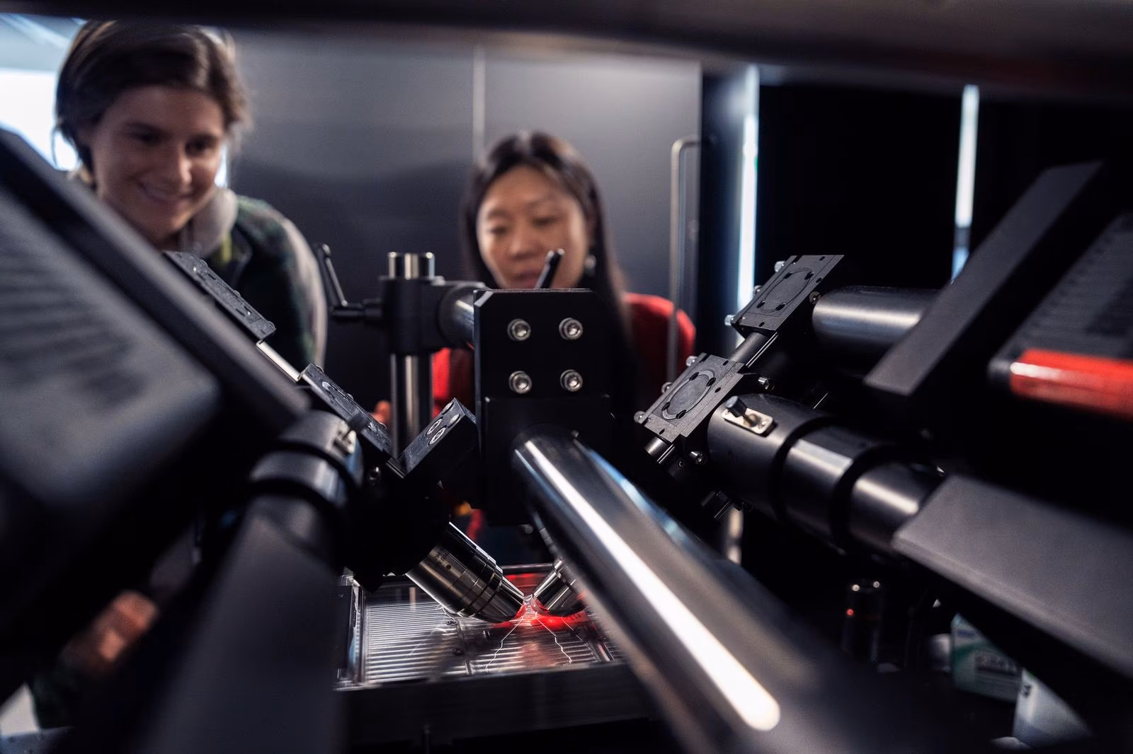
our research
our research
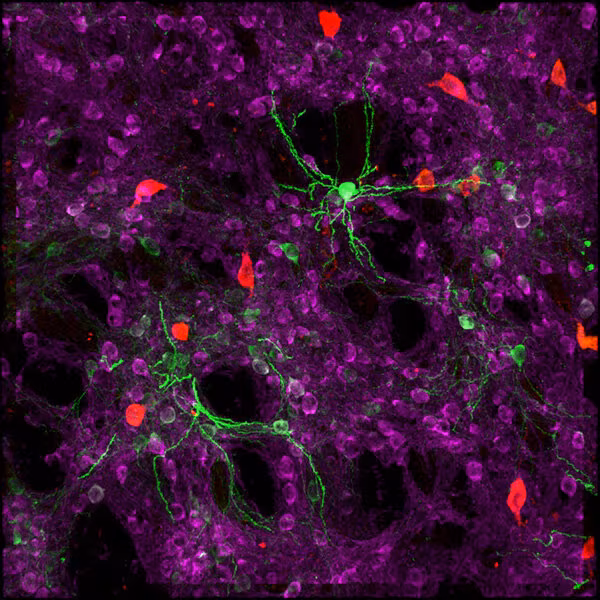
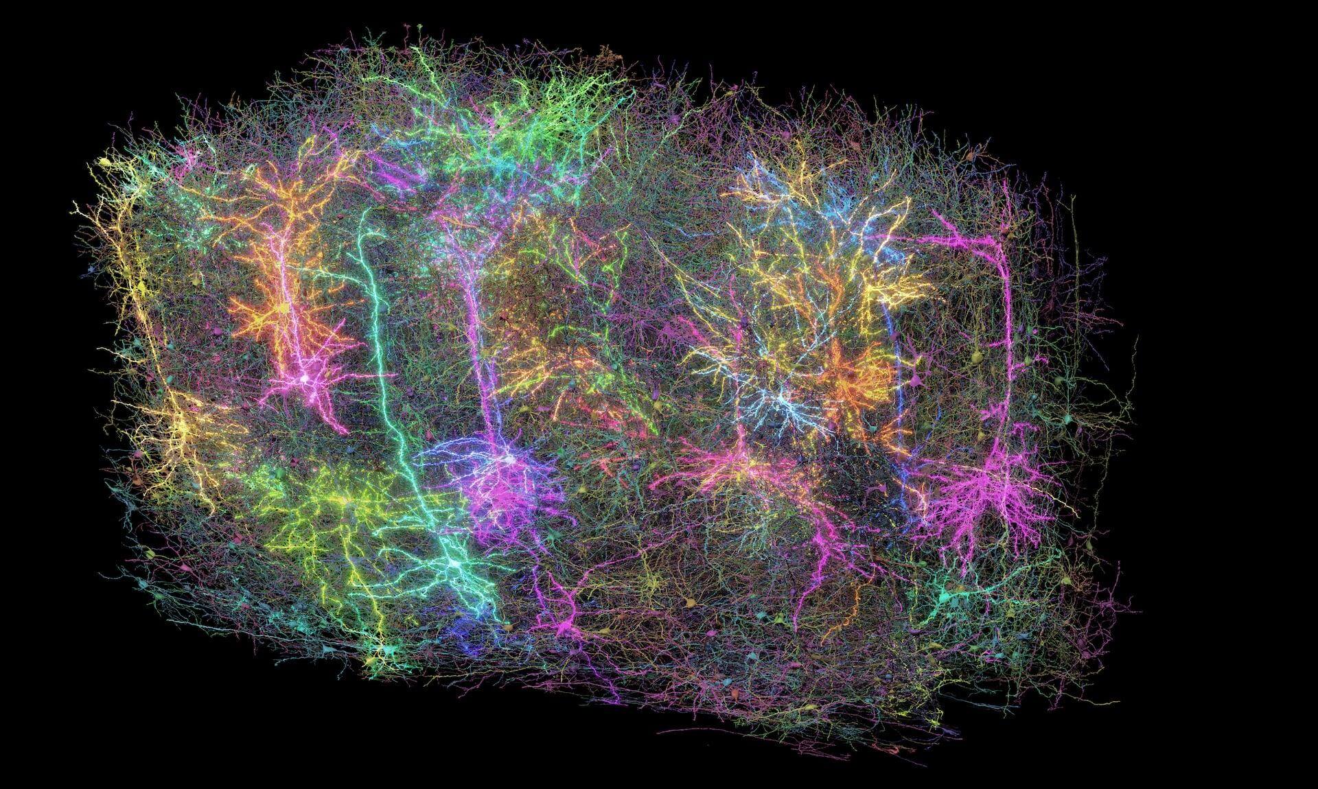
exploring the brain, cells, and immune system at the massive scale we need to transform human health
Our accelerators span scientific fields and disciplines, but they don't operate in isolation. Discoveries in one area illuminate questions in another, and our multidisciplinary teams work across boundaries. Each program runs ambitious projects with defined outcomes, generating data, tools, and knowledge shared freely with the global scientific community. This is how we accelerate discovery — not just here, but everywhere.