in this article
Neuroscientist Ueli Rutishauser, Ph.D., thought he’d uncovered a strange new phenomenon about the human brain.
He was trying to study how the brain listens to the heart — not in the figurative sense of following your heart, but by pinpointing the mysterious neurons in the brain that literally track and regulate your heartbeat.
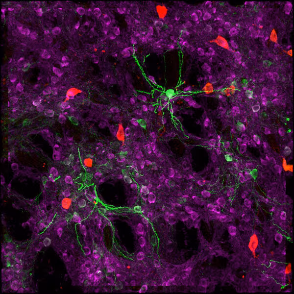
Rutishauser and his colleagues at Cedars-Sinai Medical Center in Los Angeles had precious data to try to solve that mystery: Electrical recordings taken from inside living human brains, specifically from the hippocampus, a structure deep inside the brain that’s important for memory. These data are rare, recorded from people who were either undergoing certain kinds of brain surgery or who had short-term brain implants ahead of epilepsy surgery and who agree to participate in the research studies.
When the scientists first looked at the recordings and lined them up with the patients’ heartbeats, the data jumped off the page. Almost every single neuron’s electrical activity seemed to twitch in sync with the heartbeat.
“There was this first phase of great excitement,” said Rutishauser. “We were like, ‘Oh wow, look at this. It’s such a strong effect.’ The results looked too good to be true.”
It turned out that they were.
The scientists soon realized that the neurons weren’t being affected by the heart — the tiny wires they had put into patients’ brains were. People’s brains pulse slightly as the heart beats, which, in these experiments, caused a tiny jiggle in the electrode that made the neurons’ electrical spikes look like they were changing in rhythm.
The story might have stopped there, if it weren’t for a dinner conversation Rutishauser had with Allen Institute neuroscientist Costas Anastassiou, Ph.D., that led him to realize they could use this experimental phenomenon to glean a different kind of understanding about the human brain. Ultimately, the heartbeat’s jiggle allowed the two teams of scientists to classify neurons from the patients’ brains into three different types based on their electrical signatures. The Cedars-Sinai and Allen Institute research teams published a study describing their findings in the journal Cell Reports on Tuesday.
Bridging two kinds of data about the brain
Rutishauser was part of the Next Generation Leaders Council at the Allen Institute for Brain Science, a division of the Allen Institute. This program brings early-career researchers in neuroscience to the Allen Institute for a two-way exchange of advice and expertise. At a dinner following a council meeting in Seattle three years ago, he described the heartbeat effect to Anastassiou, who leads a team of neuroscientists building and analyzing computational models of human neurons, and the two researchers set up a collaboration to delve further into the data.
“What do all these different cell types mean? How does it all come together? To address that, we have to go beyond these pieces and cells. We have to go to the intact brain.”
Allen Institute neuroscientist Costas Anastassiou, Ph.D.
Anastassiou’s computational work is part of a larger program at the Allen Institute, where researchers derive detailed data about the brain’s “parts list” from post-mortem tissue or from pieces of live brain donated by patients undergoing surgery for brain tumors or epilepsy. They use information like the genes neurons switch on, their unique electrical activity and their 3D shapes to bin these cells into different categories, or cell types.
There’s a lot they can learn about the building blocks of our brains from looking at tissue samples in the lab, but there’s a gap between that work and understanding how the different kinds of neurons produce thoughts and feelings in us, said Anastassiou, who led the Cell Reports study along with Rutishauser.
“What do all these different cell types mean? How does it all come together?” he asked. “To address that, we have to go beyond these pieces and cells. We have to go to the intact brain.”
When Rutishauser described what they had seen with the heartbeat, Anastassiou realized this could be an opportunity to bridge these two worlds: One team had detailed data about human brain cell types, but no information about what those types do in a person. The other team had recordings from living human brains, but no way of knowing which types of neurons they were listening to.
The researchers set up a collaboration and they soon realized that there were subtle differences in how different neurons’ electrical signatures changed when the electrode jiggled with the heartbeat. Anastassiou and his colleagues built a set of computational simulations of human neurons based on the Allen Institute’s human cell types data and spurred those virtual neurons to “fire” electrical spikes. They used machine learning to pair those virtual signals with the real thing, resulting in three different types of cells based on their electrical recordings.
Linking brain discoveries to cell types
The researchers have already made some insights about how these classes of neurons work in the intact brain: One of these three kinds syncs up with the patients’ theta brain waves, a slow brain wave associated with learning and plasticity in the awake brain.
“That’s not something you would necessarily see in a slice of brain in the lab,” said Cedars-Sinai postdoctoral fellow Clayton Mosher, Ph.D., who is co-first author on the study along with Allen Institute neuroscientist Yina Wei, Ph.D.
The research teams are working to define more types of human neurons using this method, and also plan to study how different neuron types switch on or off as the patients in their studies perform certain tasks, like looking at photos of family members or recall memories. Scientists have found neurons involved in such tasks — for example, a specific neuron that fires in response to pictures of Jennifer Aniston — but they don’t know what type of cell these are.
And once they can link specific cell types to memories, reactions or emotions, that opens the door to potential therapies for diseases and disorders, Rutishauser said. It’s not just that they want to understand the cell type that allows you to recognize your grandmother’s face — they also want to understand what goes wrong at the cellular level in people with dementia or other memory disorders who can no longer recognize their loved ones, and, eventually, how to correct that defect.
Citations
about the allen institute
Allen Institute is a 501(c)(3) nonprofit medical research organization dedicated to accelerating science for a healthier world. Through large-scale, multidisciplinary research initiatives, the Institute generates foundational knowledge, data, tools, and models that are shared openly with the world to advance our understanding of life and health. Founded by Jody Allen and the late Paul G. Allen, Allen Institute is supported primarily by the Fund for Science and Technology.