in this article
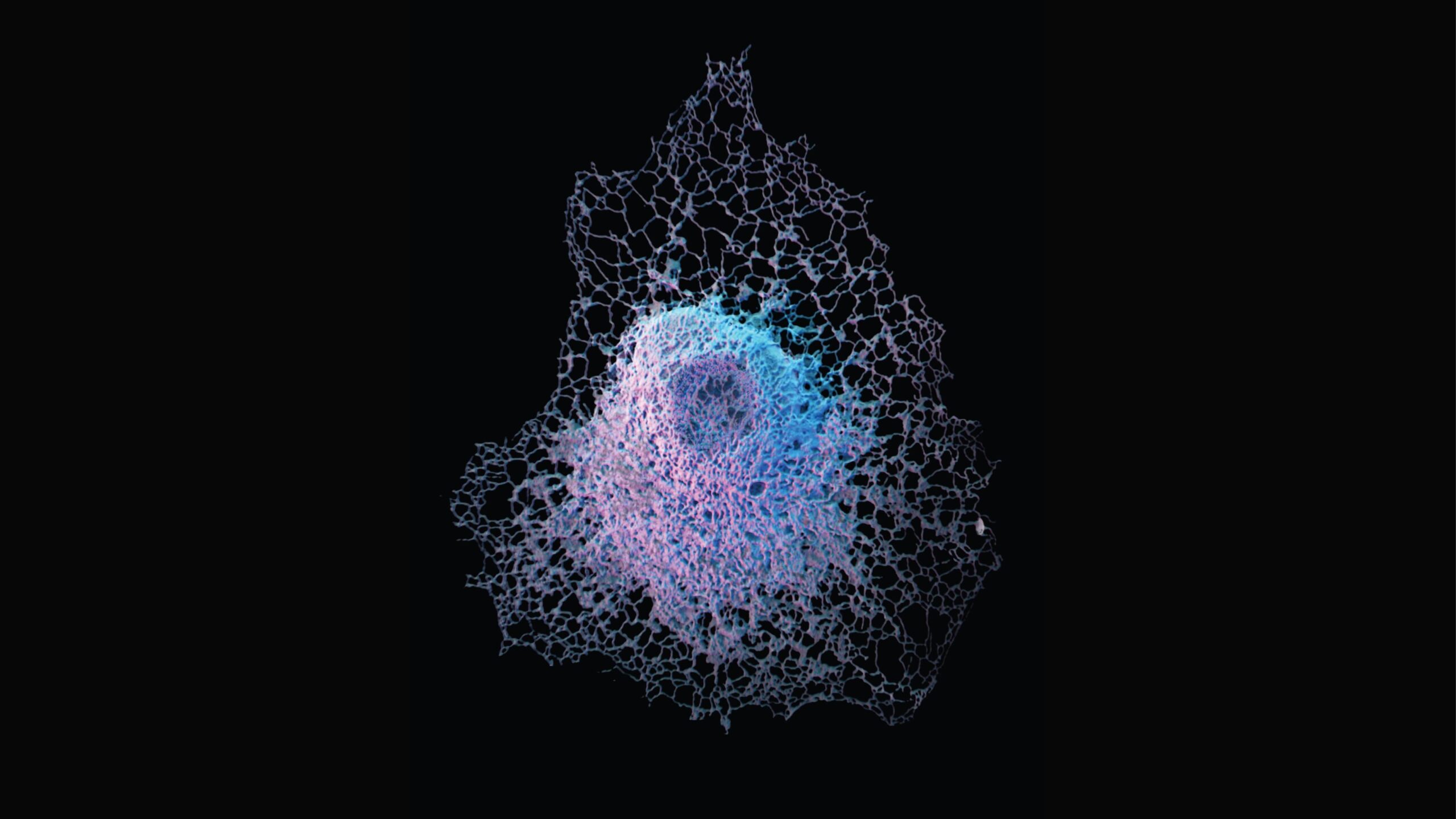
The insides of your cells are kind of a mess. Far from the neatly packaged bubble-like structures your high school biology textbook illustrations showed, cellular structures — also known as organelles — are often branching, jumbled, and crowded together. Andrew Moore, Ph.D., a biologist at HHMI’s Janelia Research Campus, is studying one of these organelles, shown in this image, known as the endoplasmic reticulum or ER. This large, multi-folded structure surrounds the cell’s nucleus (the storage center for your chromosomes in each cell) and is the site of protein production and folding. Moore wants to understand how the ER gets and keeps its complicated shape, and how it interacts with the scaffolding structures of the cell. To create this image that shows the details of the ER’s outer membrane, Moore used a graphics tool built by researchers at the Allen Institute for Cell Science known as AGAVE. The tool borrows techniques from animation studios to create realistic light sources and shadows in a flat image, making it look as realistic and 3D as possible.
Citations
about the allen institute
Allen Institute is a 501(c)(3) nonprofit medical research organization dedicated to accelerating science for a healthier world. Through large-scale, multidisciplinary research initiatives, the Institute generates foundational knowledge, data, tools, and models that are shared openly with the world to advance our understanding of life and health. Founded by Jody Allen and the late Paul G. Allen, Allen Institute is supported primarily by the Fund for Science and Technology.