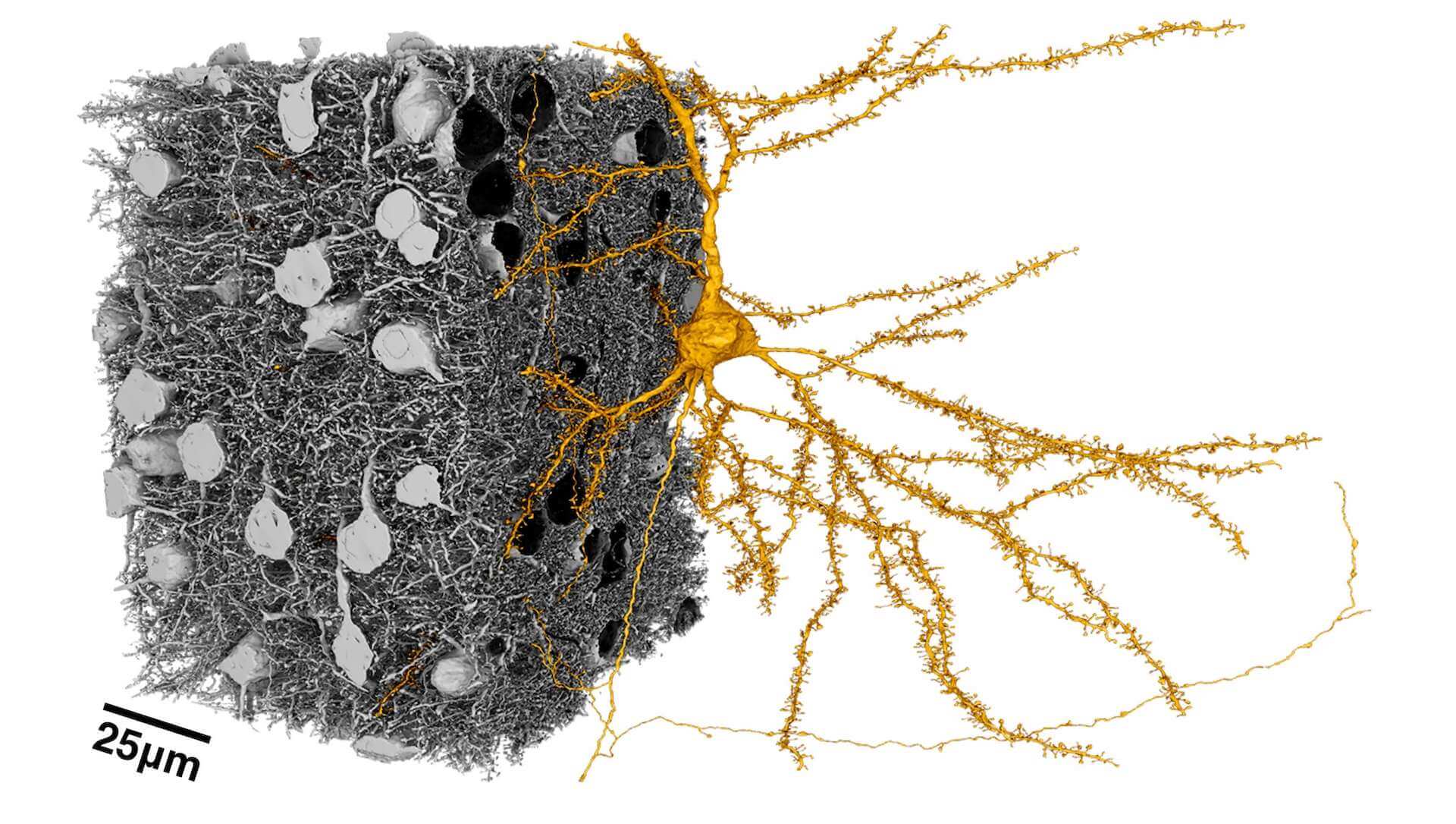

Neuroscientists are using a method known as electron microscopy to view neurons in a completely new way. The special machines, which use electrons instead of light waves to visualize structures, revealing incredibly fine levels of detail about an entire microscopic landscape — in this case, the densely packed topography of neurons and other cells that make up the brain’s physical matter. In a five-year collaboration between the Allen Institute, Princeton University and Baylor College of Medicine, funded and coordinated by the Intelligence Advanced Research Projects Activity, or IARPA, scientists and engineers have mapped fine structures, and connections and functional properties in a tiny piece of the mouse brain from the visual neocortex, the part of the brain that processes what we see. The research teams recently published an article in the journal Cell describing some of these neurons, many of which are a kind known as a pyramidal cell, shown here in gold in an image from the collaborative study. Pyramidal cells have a large, pyramid shaped body and send excitatory signals, meaning they connect with other neurons to switch on their activity. By studying these cells’ activity in a living mouse before imaging them under the microscope, the scientists have been able to capture important information about how pyramidal cells’ activity correlates with their detailed structure and connections.
Citations
about the allen institute
Allen Institute is a 501(c)(3) nonprofit medical research organization dedicated to accelerating science for a healthier world. Through large-scale, multidisciplinary research initiatives, the Institute generates foundational knowledge, data, tools, and models that are shared openly with the world to advance our understanding of life and health. Founded by Jody Allen and the late Paul G. Allen, Allen Institute is supported primarily by the Fund for Science and Technology.