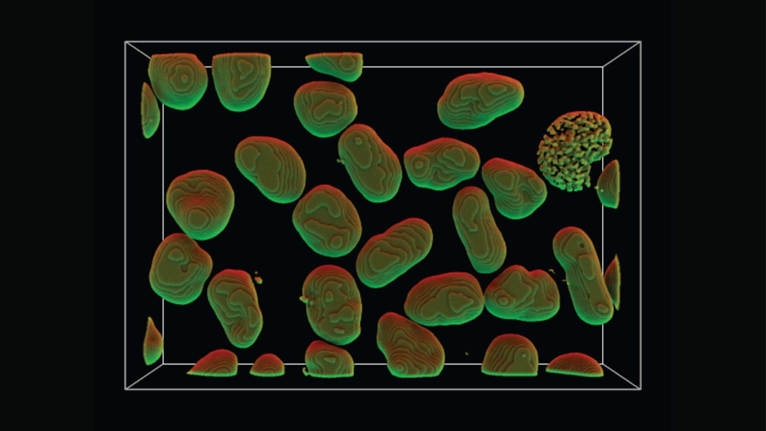
Researchers at the Allen Institute for Cell Science want to know what a normal cell looks like. It might sound like a simple research question, but after analyzing images of hundreds of thousands of human stem cells with different tags to light up different parts of the cell, the scientists are coming to understand just what a huge range of normal exists among even the same kind of human cell growing in a dish. In the image above, a protein known as h4B is genetically engineered to light up under the microscope. h4B is part of a structure that organizes DNA inside the cell’s nucleus; this image thus represents the overall shape of human DNA and its associated proteins in each cell. The image was processed to find the clean edges of the structures and then further modified using an in-house graphics tool known as AGAVE, which creates realistic-looking light sources. Here, software engineer Dan Toloudis used the tool to “shine” green and red light on the DNA structures. —Rachel Tompa, Ph.D.
Microscopic viewpoints, computer-generated models, intricate tracings and more — see a new side of science with SciShots.
Citations
about the allen institute
Allen Institute is a 501(c)(3) nonprofit medical research organization dedicated to accelerating science for a healthier world. Through large-scale, multidisciplinary research initiatives, the Institute generates foundational knowledge, data, tools, and models that are shared openly with the world to advance our understanding of life and health. Founded by Jody Allen and the late Paul G. Allen, Allen Institute is supported primarily by the Fund for Science and Technology.