in this article
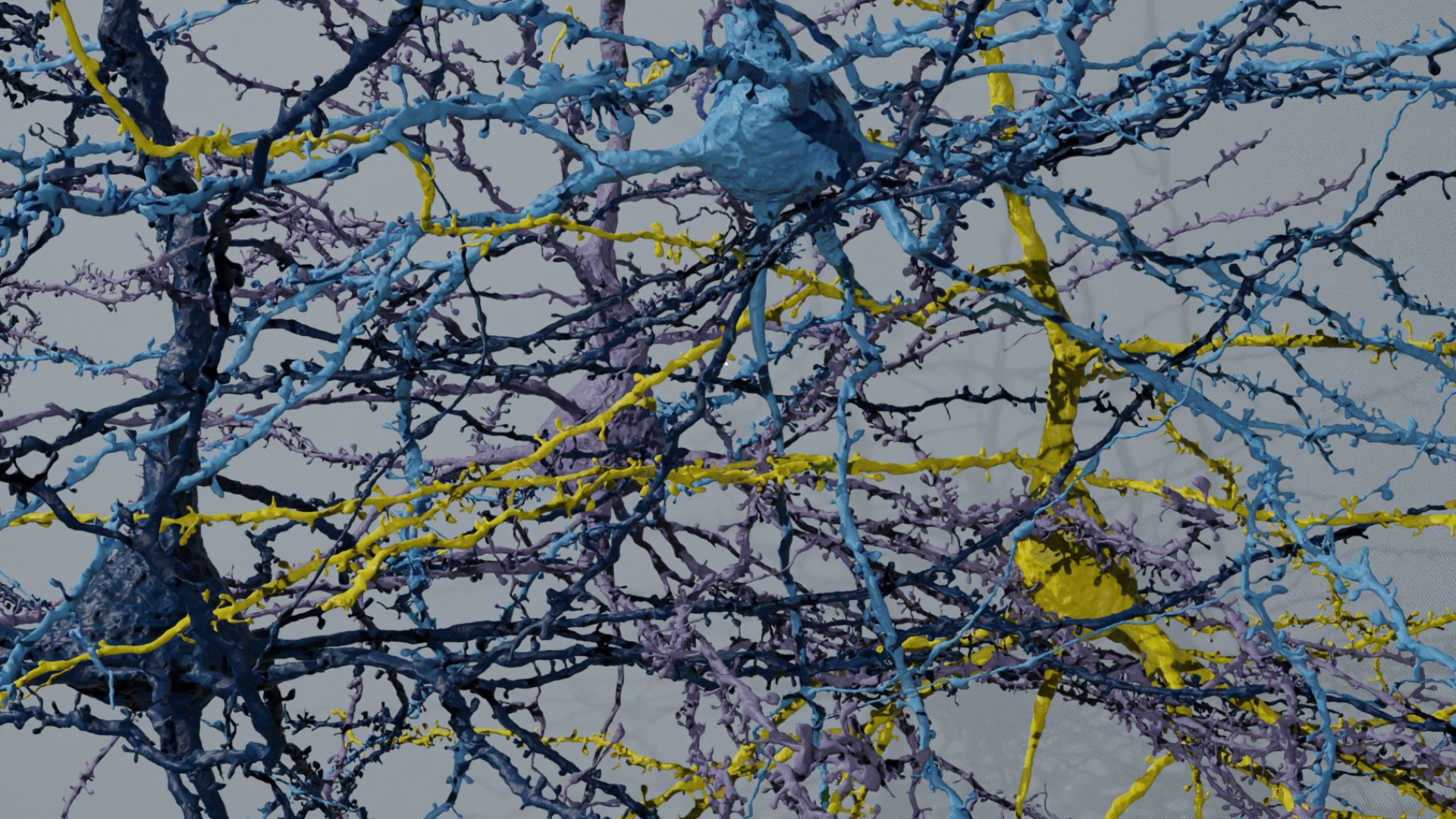
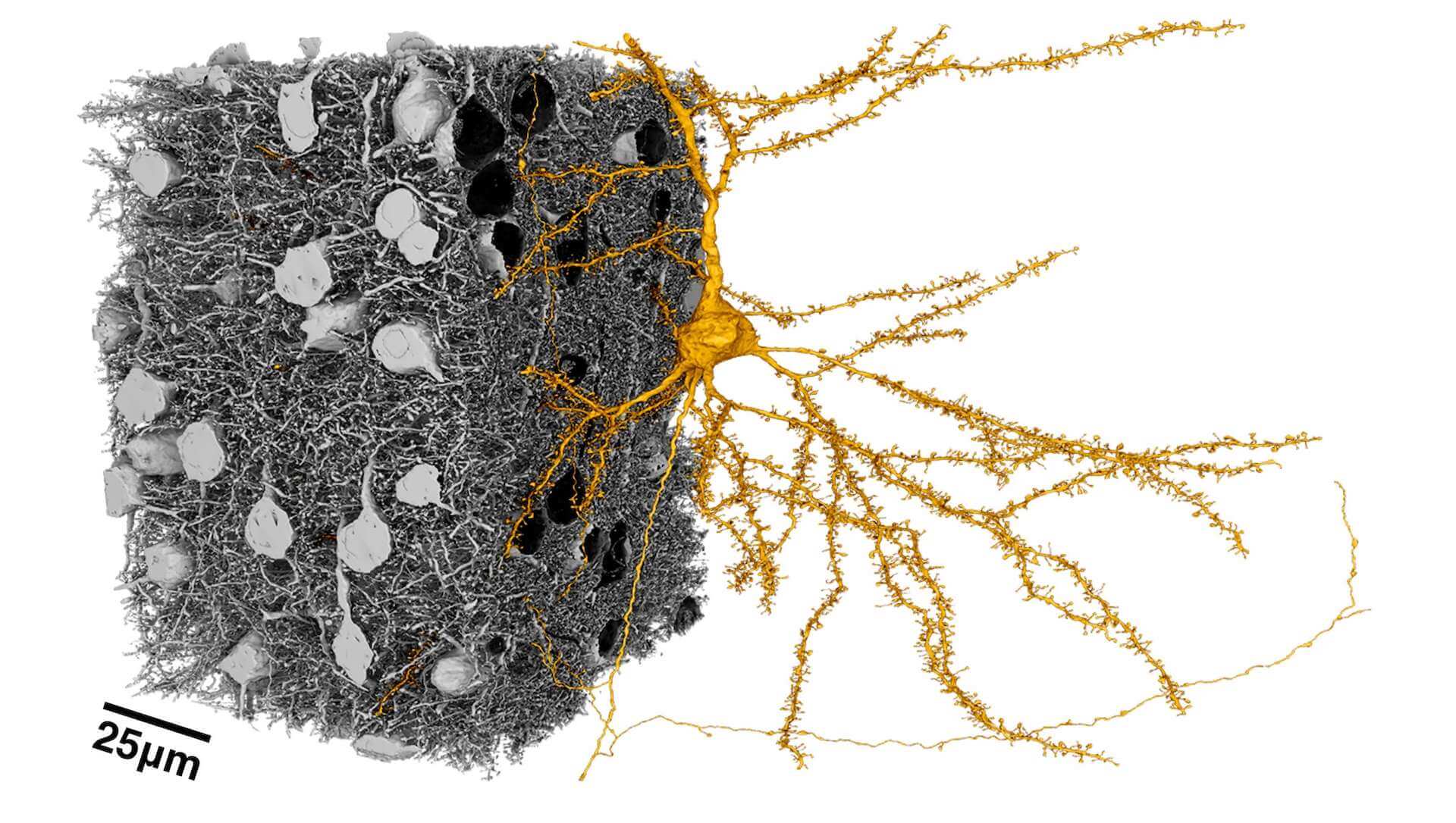
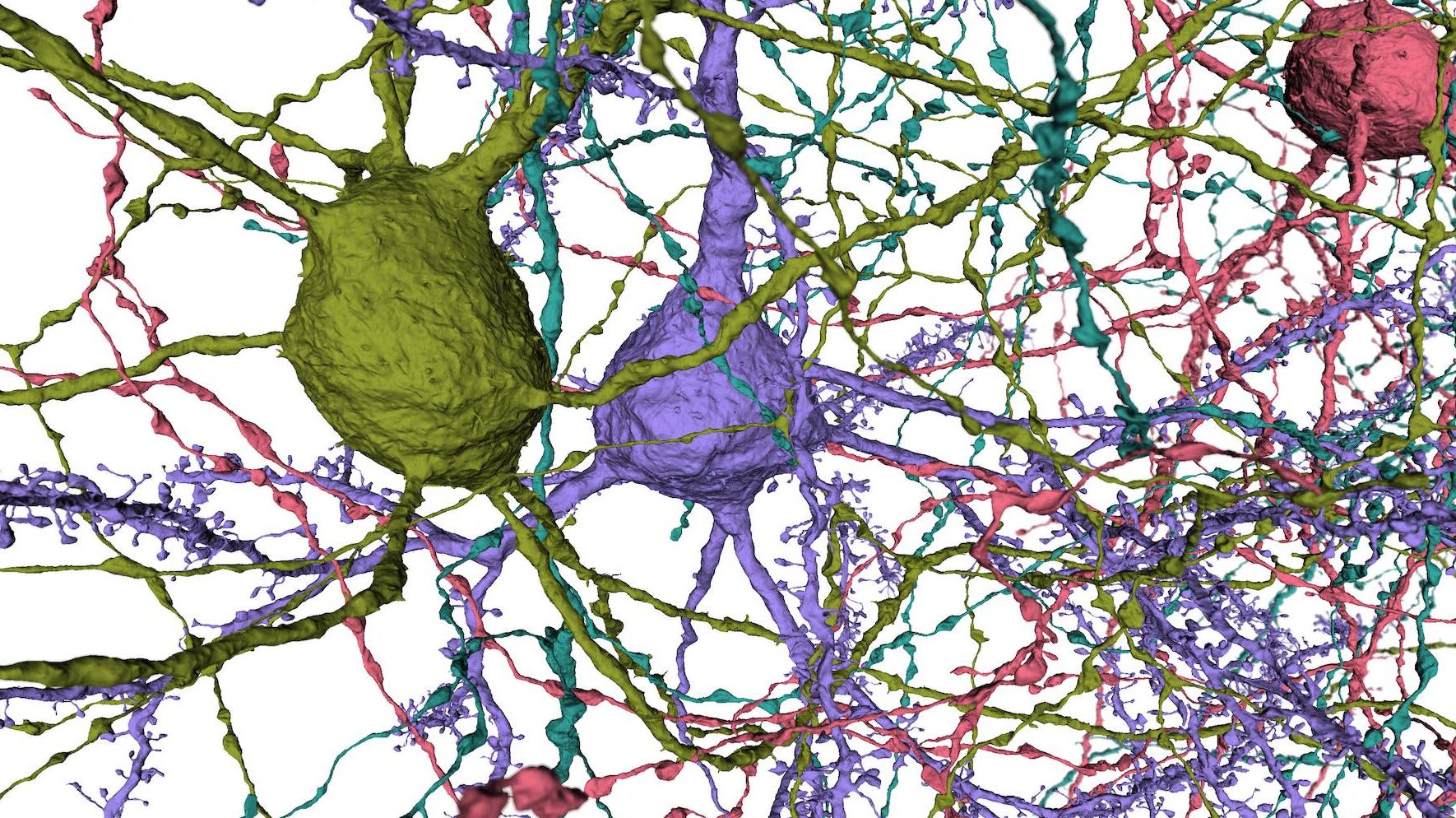
To truly understand the brain, we need a roadmap of how it’s wired. The gold standard for studying the cellular architecture and then connectivity of brain cells is what scientists call serial-section electron microscopy. This type of imaging requires cutting brain samples into ultra-thin slices and taking pictures of those slices with an electron microscope. Those images can then be sandwiched together to create a three-dimensional picture of brain cells and their connections.
For decades, this work has been painstakingly done by hand. To greatly speed the process, researchers at the Allen Institute built a high-throughput electron microscopy pipeline called piTEAM: parallel imaging transmission electron automated microscopes. Details of this pipeline published today in the journal Nature Communications.
In 2016, with the support of IARPA, the Allen Institute for Brain Science joined a collaboration with Baylor College of Medicine and Princeton University to build high-resolution roadmap of wiring in the brain. The Allen Institute’s role was to do something that had never been done before: cut a one cubic millimeter section of brain cortex into ~25,000 ultra-thin slices, take ~125,000,000 pictures of those slices and assemble them into a 3D volume containing ~100,000 cells, 2.5 miles of wiring and 1,000,000,000 synaptic connections.
The piTEAM pipeline includes highly complex automated cutting machines and microscopes that can run 24/7 with minimal human intervention. This approach is not unlike the fly-by-wire avionics in modern passenger jets that allow digital control of flight systems.
In 2018, the Allen Institute team imaged more than 26,500 sections of mouse cortex, across five microscopes, for nearly six months. In the end, they collected ~125,000,000 images of the brain at synapse resolution. All of those images totaled ~2 petabytes; enough data to fill up nearly 4,000 high-capacity iPhones. The result of these data will be the most complete wiring diagram of a mammal’s cortex to date.
Citations
about the allen institute
The Allen Institute is an independent, 501(c)(3) nonprofit research organization founded by philanthropist and visionary, the late Paul G. Allen. The Allen Institute is dedicated to answering some of the biggest questions in bioscience and accelerating research worldwide. The Institute is a recognized leader in large-scale research with a commitment to an open science model. For more information, visit alleninstitute.org.