in this article
SciShots: Clusters of Alzheimer’s cells
New data explores the cellular landscape of Alzheimer’s disease
08.05.2022
1 min read
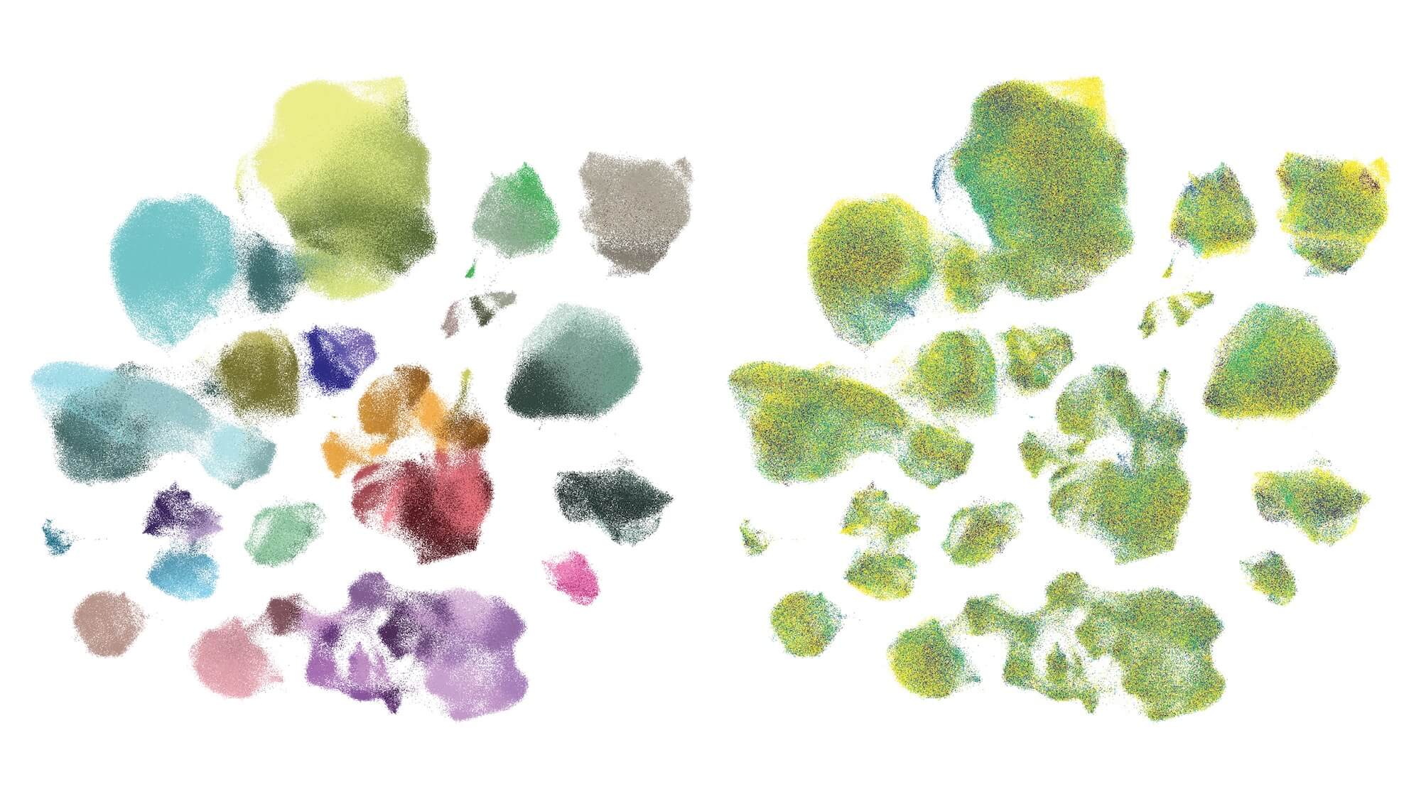
A newly released dataset of 1.2M cells from 84 brain donors across the spectrum of Alzheimer’s shines a light on the neurodegenerative disease’s cellular roots. Produced by the Seattle Alzheimer’s Disease Brain Cell Atlas consortium, an NIA-funded collaboration of Seattle-area scientists headquartered at the Allen Institute, the data capture several different aspects of the cellular and molecular underpinnings of this disease. The image above from the Allen Institute team shows brain cells from all 84 donors clustered and colored by the type of cell (left) or by the level of Alzheimer’s-related proteins present (right, yellow is high and blue is low). The scientists used genes switched on in individual brain cells to categorize them into discrete types, methods first used to understand the basic cellular building blocks of a healthy brain and which are now being applied to study disease at a new level of resolution. —Rachel Tompa, Ph.D.
Microscopic viewpoints, computer-generated models, intricate tracings and more — see a new side of science with SciShots.
About the author: Rachel Tompa
Rachel Tompa is a science and health writer and editor. A former molecular biologist, she’s been telling science stories since 2007 and has covered the gamut of science topics, including the microbiome, the human brain, pregnancy, evolution, science policy and infectious disease. During her tenure as Senior Editor at the Allen Institute, Rachel wrote stories and created podcast episodes covering all the Institute’s scientific divisions.
Get in touch at press@alleninstitute.org.
Citations
about the allen institute
Allen Institute is a 501(c)(3) nonprofit medical research organization dedicated to accelerating science for a healthier world. Through large-scale, multidisciplinary research initiatives, the Institute generates foundational knowledge, data, tools, and models that are shared openly with the world to advance our understanding of life and health. Founded by Jody Allen and the late Paul G. Allen, Allen Institute is supported primarily by the Fund for Science and Technology.