in this article
As part of the suite of tools publicly available on brain-map.org, the Allen Institute for Brain Science has launched the Allen Brain Observatory: the first tool of its kind to provide a highly standardized survey of cellular-level activity in the mouse visual system. With the data and tools in this resource, researchers around the world are empowered to investigate how circuits in the mouse brain coordinate while the mouse performs visual tasks, taking in and processing a wide range of visual stimuli.
Allen Brain Observatory: Visualizing the brain in action
“The Allen Institute is known for our atlases—deep, high quality data sets revealing where genes are expressed and how cells and connections are arranged in the mouse and human brain,” says Allan Jones, Ph.D., CEO of the Allen Institute. “With the Allen Brain Observatory, we’ve taken an important leap into measuring natural brain activity as it is actually happening.”
“The Allen Brain Observatory is a stunning window into the visual brain of the mouse,” says Christof Koch, Ph.D., President and Chief Scientific Officer of the Allen Institute for Brain Science. “No one has ever taken this kind of standardized approach to surveying the active brain at cellular resolution in order to measure how the brain processes information in real time. This is a milestone in our quest to decode how the brain’s computations give rise to perception, behavior, and consciousness. Just like in astronomy, modelers and theoreticians worldwide can now study this wealth of data using their own analysis tools.”
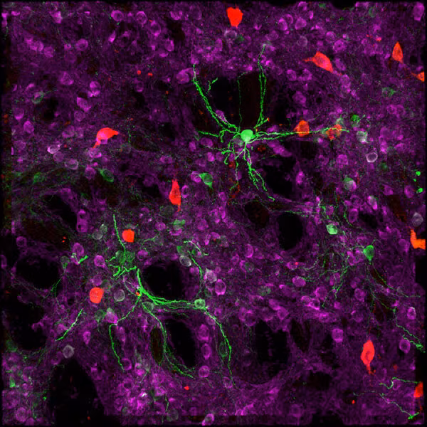
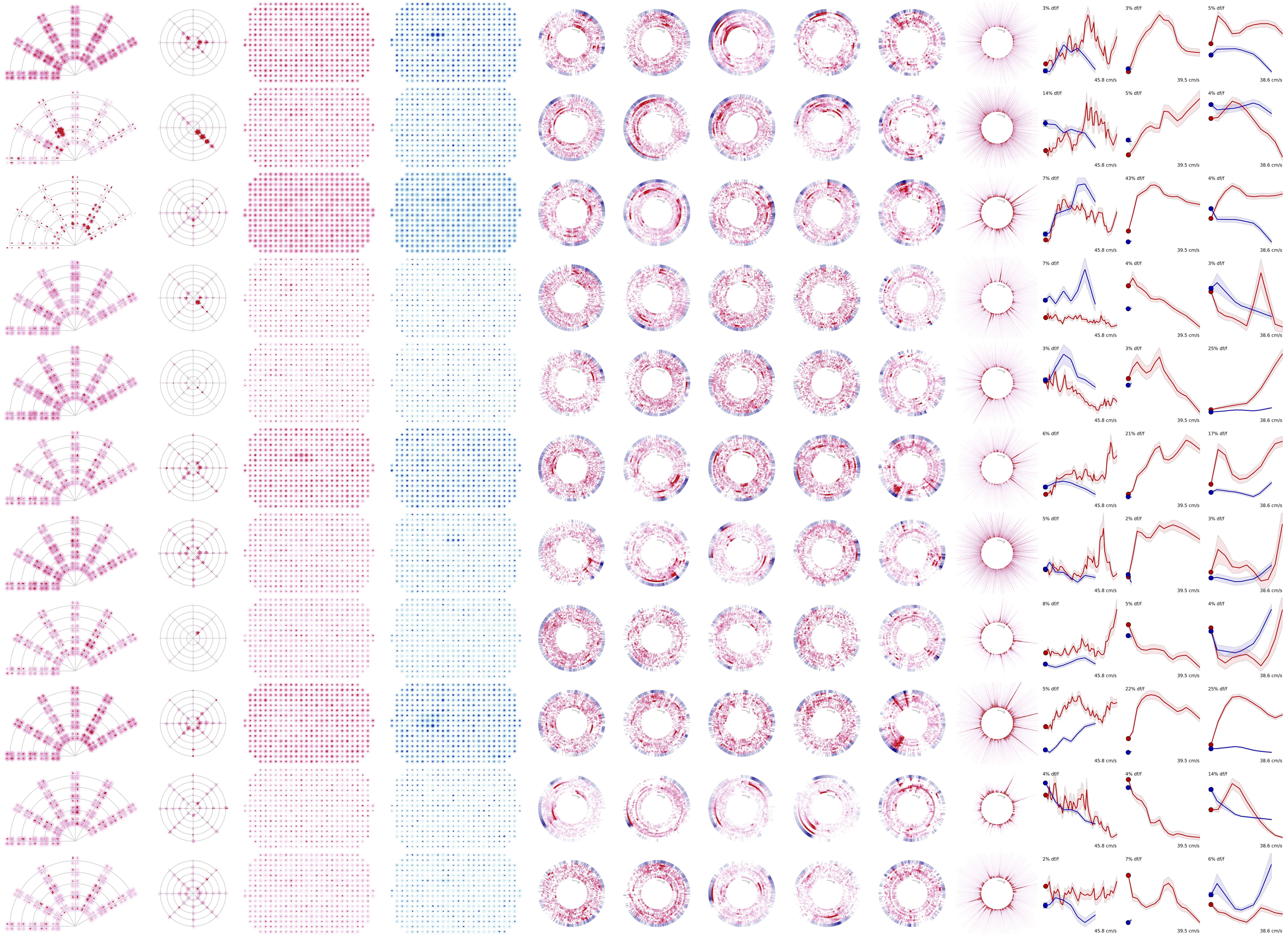
The first data in the Allen Brain Observatory survey four areas in the mouse visual cortex at multiple depths, sampling more than 18,000 neurons in total. The mice were presented with a large variety of visual stimuli to determine the “tuning,” or preference, of each individual cell to visual features like motion and shape orientation, as well as complex images like natural scenes and movies that reveal integrative dynamics of visual processing.
The data from thousands of individual cells and populations of cells are presented in a novel visualization format through the Allen Brain Atlas data portal, and are accompanied by analysis tools and access to all raw data, which allows scientists to deeply explore the rules that govern how networks of cells in the visual cortex communicate.
More than 100 people at the Allen Institute were involved in the creation of the Allen Brain Observatory, from animal care technicians to neuroscientists, engineers, microscopy experts, optical physiologists, physicists, mathematicians and computer scientists.
Learn more about the Allen Brain Observatory in our press release, or visit the Allen Brain Observatory at brain-map.org.
GO TO THE ALLEN BRAIN OBSERVATORY
Press Coverage
- Nature – Brain-data gold mine could reveal how neurons compute
- WIRED – 35 Mice Watched the Cult Film Touch of Evil for Science
- Forbes – What Paul Allen, Orson Welles And A Mouse Are Teaching Us About Our Brains
- NPR – A Mouse Watches Film Noir And Offers Clues To Human Consciousness
- STAT: Boston Globe – This team is showing movies to mice in hopes of unlocking the brain’s secrets
- The Atlantic – The Many Ways to Map the Brain
- Seattle Times – What happens if you show a mouse a movie: Allen Institute opens new window into living brain
- GeekWire – Allen Brain Observatory peers into the minds of mice as they watch movies
- Tech Insider – A lab founded by a tech billionaire just unveiled a major leap forward in cracking your brain’s code
- The Scientist – Allen Institute Launches Brain Observatory
- International Business Times – Mapping The Brain: Allen Institute Launches Observatory To Study Perception And Cognition
- Tech Times – Here’s What Scientists Uncovered By Letting Mice Watch Film Noir
Citations
about the allen institute
Allen Institute is a 501(c)(3) nonprofit medical research organization dedicated to accelerating science for a healthier world. Through large-scale, multidisciplinary research initiatives, the Institute generates foundational knowledge, data, tools, and models that are shared openly with the world to advance our understanding of life and health. Founded by Jody Allen and the late Paul G. Allen, Allen Institute is supported primarily by the Fund for Science and Technology.