Attempting the impossible: A 20-year journey to learn the language of the brain
by Peter Kim
Video credit: Tyler Sloan, Ph.D., Quorumetrix Studio
Explore the connections of the brain – MICrONS neuroscience data insights.

“It is no use asking for the impossible, such as, say, the exact wiring diagram for a cubic millimeter of brain tissue and the way all its neurons are firing.”
— Francis Crick, 1916 – 2004
That quote first appeared in a 1979 essay in Scientific American.
In the early 80s, Clay Reid, M.D., Ph.D., was finishing college at Yale and read that essay by famed molecular biologist Francis Crick. The article inspired many young scientists and fueled many of the 21st Century’s advances in studying the function and structure of the brain. The words lit a fire in Reid, and others, who became interested in the brain and the spectacular complexity of its wiring.
“The whole story of that quote makes me happy. Here is the Scientific American from 1979. I got a copy of this and was just astounded to read…”
In 2025, 46 years after Crick’s quote, a global team of over 150 scientists and researchers completed the largest and most complex digital reconstruction of one cubic millimeter of the mouse visual cortex—a massive leap forward in accomplishing the “impossible” task Crick first described.
The Machine Intelligence from Cortical Networks (MICrONS) Project maps the neurons, their electrical activity, and their connection patterns in a single, massive wiring diagram rich with insight.
The seed for this incredible undertaking was planted back in 1979, with that special issue of Scientific American that inspired many scientists. Reid and his early team of researchers at Harvard were among the early founders of the field of electron microscopy (EM) connectomics, which is the foundation for the MICrONS Project.
“The project started almost 20 years ago and it was very soon after the first paper in what became the field of EM connectomics and a student and I just decided this is the time, let’s start reconstructing neural circuits with electron microscopy.”
In 2012, Clay left Harvard Medical School, where he was a professor, to pursue the “impossible” at the Allen Institute—mapping the connections together with the functional properties of a one cubic millimeter portion of the mouse brain.
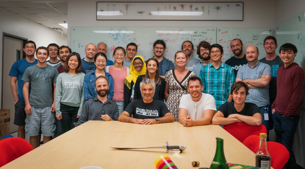
He assembled the team that would lead and manage the completion of this massive moonshot project—the most challenging neuroscience experiment ever attempted.
The team included key scientists like Nuno da Costa, Ph.D., as experimental lead
… and Forrest Collman, Ph.D., data and technology lead, along with scientists at Princeton, Baylor College of Medicine and many others. Over 150 scientists and researchers were a part of the project—a monumental team effort involving multiple labs and institutions.
Analysis of the wiring diagram resulted in ten studies that were published in the Nature family of journals.
By the Numbers
petabytes of data
Scientists mapped around 4 kilometers of axons. That’s the distance from the Allen Institute in Seattle to Lumen Field.
The team originally at Baylor College of Medicine (now at Stanford University), in the lab of Andreas Tolias, Ph.D., collected the functional data, recording the electrical activity from tens of thousands of neurons using calcium imaging.
For this extraordinary task, researchers showed mice visual stimuli, like the video currently running in the background, and recorded the resulting brain activity from individual neurons in the visual cortex.
Using electron microscopy, scientists at the Allen Institute then took around 95 million high-resolution images of that same section of the mouse visual cortex researchers at Baylor analyzed.
Scientists in the lab of Sebastien Seung, Ph.D., from Princeton University, then assembled and aligned the images into a 3D reconstruction revealing unprecedented detail and connection.
“MICrONS will be remembered as the beginning of a digital transformation of neuroscience. In this new era, computational neuroscience is moving from the fringes to the center of our discipline, and connectomes are becoming the foundation.”
— Sebastian Seung, Ph.D., Princeton Neuroscience Institute
Combined with the electrical activity data from Baylor, the MICrONS wiring diagram reveals form and function and begins to reveal the language and logic brain cells use to communicate—the elusive algorithms of the cortex.
It is a powerful research tool, openly available for the global scientific community, that could lead to new treatments for brain disorders.

The importance of combining form and function
The collaborative, global effort was made possible by support from the Intelligence Advanced Research Projects Activity (IARPA) and National Institutes of Health’s Brain Research Through Advancing Innovative Neurotechnologies® Initiative, or The BRAIN Initiative®.

“The BRAIN Initiative plays a critical role in bringing together scientists from various disciplines to perform complex and challenging research that cannot be achieved in isolation. Basic science building blocks, like how the brain is wired, are the foundation we need to better understand brain injury and disease, to bring treatments and cures closer to clinical use.”
— John Ngai, Director, NIH BRAIN Initiative
The MICrONS Project began with words on a page in 1979. And those words are now helping scientists around the world learn the language of the brain.

Produced By
Brian Cama / Allen Institute
Jenny Burns / Allen Institute
Liz Dueweke / Allen Institute
Peter Kim / Allen Institute
Scientific Collaborators
Princeton University
Baylor College of Medicine
Special Thanks
Tyler Sloan, Quorumetrix Studio