impact
about
our story
explore our impact
careers and opportunities
join us
open science
sharing science to speed discovery
open science week
celebrating open science
team science
people & teams
discovery is a team effort
allenites
the people who make our work possible
advisors
their input shapes our science
board of directors
they help us fulfill our mission
shanahan foundation fellowship
at the interface of data and neuroscience
next generation leaders
fostering emerging leaders in bioscience
research
overview
our approach
science at the scale of greatest impact
publications
explore our publications
open science
sharing science to speed discovery
science accelerators
brain science
building a blueprint of the brain
cell science
understanding how cells become organs
neural dynamics
revealing the brain’s hidden algorithms
immunology
creating the ultimate immune system reference
synthetic biology
seattle hub for synthetic biology
education
overview
science education
science is for everyone
open science
sharing science speeds discovery
engagement
education resources
real science. real skills.
field trips
experience science where it happens
educator development
empowering educators
news
overview
all news
explore the latest news
podcast
the human stories behind discovery
sign up for our newsletter
stay connected to our science
events
overview
all events
public engagement, workshops, seminars and more
conferences
connect with us
science resources
science resources
allencell.org
allenimmunology.org
allenneuraldynamics.org
brain-bican.org
brain-map.org
microns-explorer.org
impact
back to menu
team science
our story
explore our impact
careers and opportunities
join us
open science
sharing science to speed discovery
open science week
celebrating open science
team science
people & teams
discovery is a team effort
allenites
the people who make our work possible
advisors
their input shapes our science
board of directors
they help us fulfill our mission
shanahan foundation fellowship
at the interface of data and neuroscience
next generation leaders
fostering emerging leaders in bioscience
research
back to menu
overview
our approach
science at the scale of greatest impact
publications
explore our publications
open science
sharing science to speed discovery
science accelerators
brain science
building a blueprint of the brain
cell science
understanding how cells become organs
neural dynamics
revealing the brain’s hidden algorithms
immunology
creating the ultimate immune system reference
synthetic biology
seattle hub for synthetic biology
education
back to menu
overview
science education
science is for everyone
open science
sharing science speeds discovery
engagement
education resources
real science. real skills.
field trips
experience science where it happens
educator development
empowering educators
news
back to menu
overview
all news
explore the latest news
podcast
the human stories behind discovery
newsletter
stay connected to our science
events
back to menu
overview
all events
public engagement, workshops, seminars and more
conferences
connect with us
science resources
back to menu
science resources
allencell.org
allenimmunology.org
allenneuraldynamics.org
brain-bican.org
brain-map.org
microns-explorer.org
search
research
brain science

connectomics

mapping the nervous system at cellular and synaptic resolution within and across brain regions
Colorful neon neural network visualization with branching lines on black background

goals and approach

The Connectomics team at the Allen Institute's brain science accelerator uses large scale anatomical methods to measure connectivity within the brain at a very large scale with single–cell resolution. There are two major projects within the department that operate at different scales. Electron microscopy (EM) connectomics attempts to measure connectivity and morphology of cells with single synapse resolution across millimeters of brain. Axonal connectomics uses light microscopy methods to reconstruct large caliber single axons as they course the brain’s white matter, at the scale of centimeter, in primate and human brains. Both projects also characterize and classify cell types based on their morphology and work on integrating these morphological classifications with other modalities such as transcriptomics and single-cell physiology, both in vivo and in vitro.

Team of researchers posing with brain scan visualization on large display monitor in laboratory
3D illustration of interconnected branching neural networks with golden yellow and blue gradient colors
A rendering of reconstructions of layer 5 thick tufted neurons from the visual cortex of a mouse. Data from microns-explorer.org
‍
Project leads:
Nuno da Costa, Forrest Collman & Clay Reid
‍
‍Team members: Agnes Bodor, Derrick Brittain, Steven Cook, Bethanny Danskin, Cameron Devine, Chris Jordan, Cheryl Lea, Melissa Lerch, Xiaoyu Lu, Sid Rath, Ben Pedigo, Jenna Schardt, Casey Schneider-Mizell, Rachael Swanstrom, Marc Takeno, Russel Torres, Keith Wiley, Wan-qing Yu, Chi Zhang, Kim Gruver

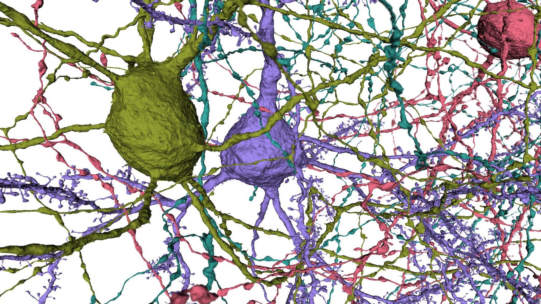
electron microscopy connectomics

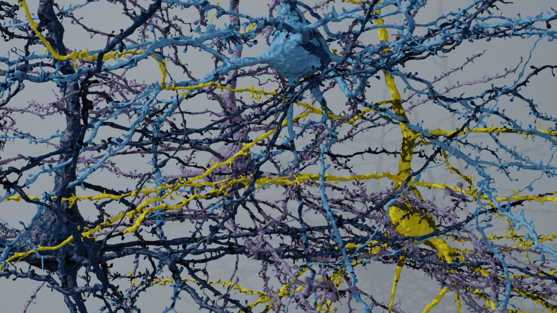
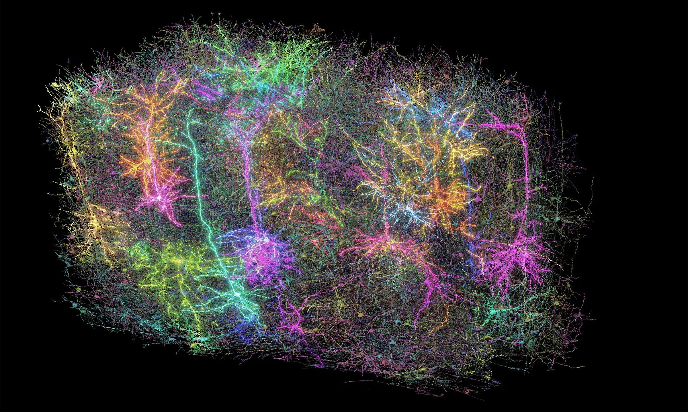
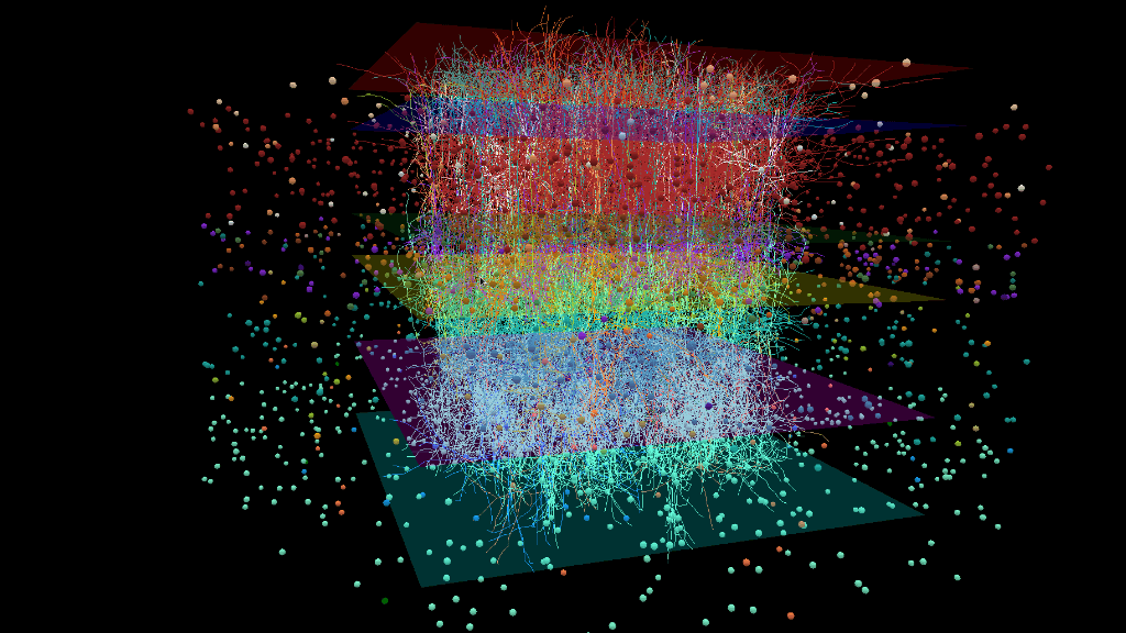
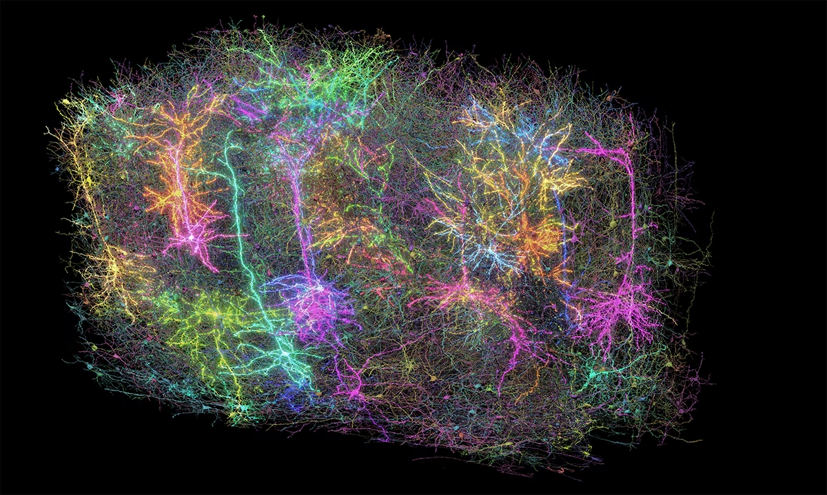
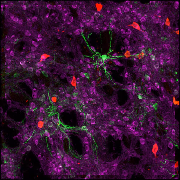
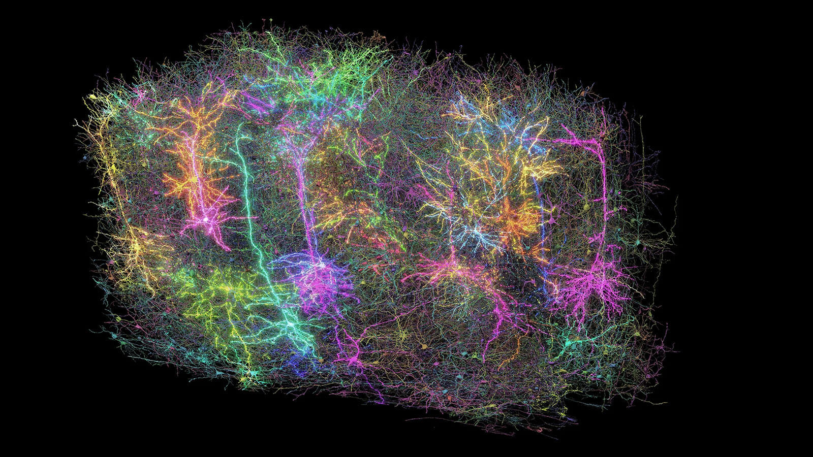
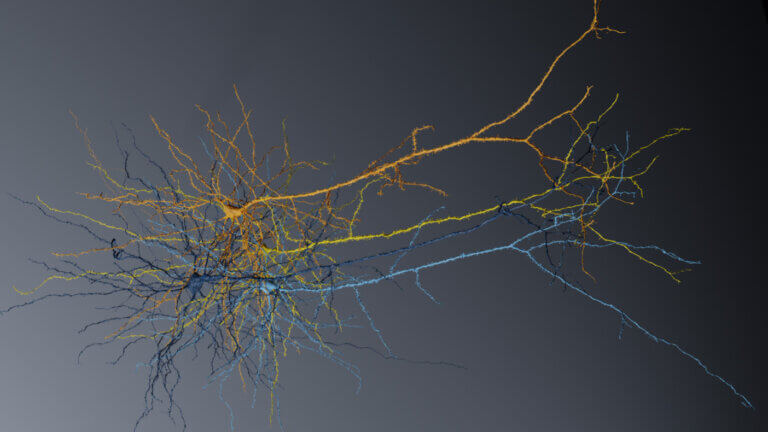
In order to reconstruct neural circuits at a large scale with synaptic resolution, electron microscopy offers unique advantages in capturing the detailed morphology of all individual neurons and non-neuronal cells within a volume. By applying high throughput sectioning, imaging, image processing, and automated segmentation, our team contributes to large-scale reconstructions of neural circuits. We have published work primarily on the reconstruction of visual circuits of the mouse, but have future looking projects moving toward reconstructions of motor circuits in mouse and monkey, and basal ganglia circuits of the mouse. We are also leading one of the centers of the larger NIH BRAIN CONNECTS effort to scale up pipelines to capture the synaptic connectivity of the entire whole mouse. Our team consists of an interdisciplinary group that combines expertise in electron microscopy, neuroscience, computer science, mechanical engineering and program management to execute this complex pipeline.
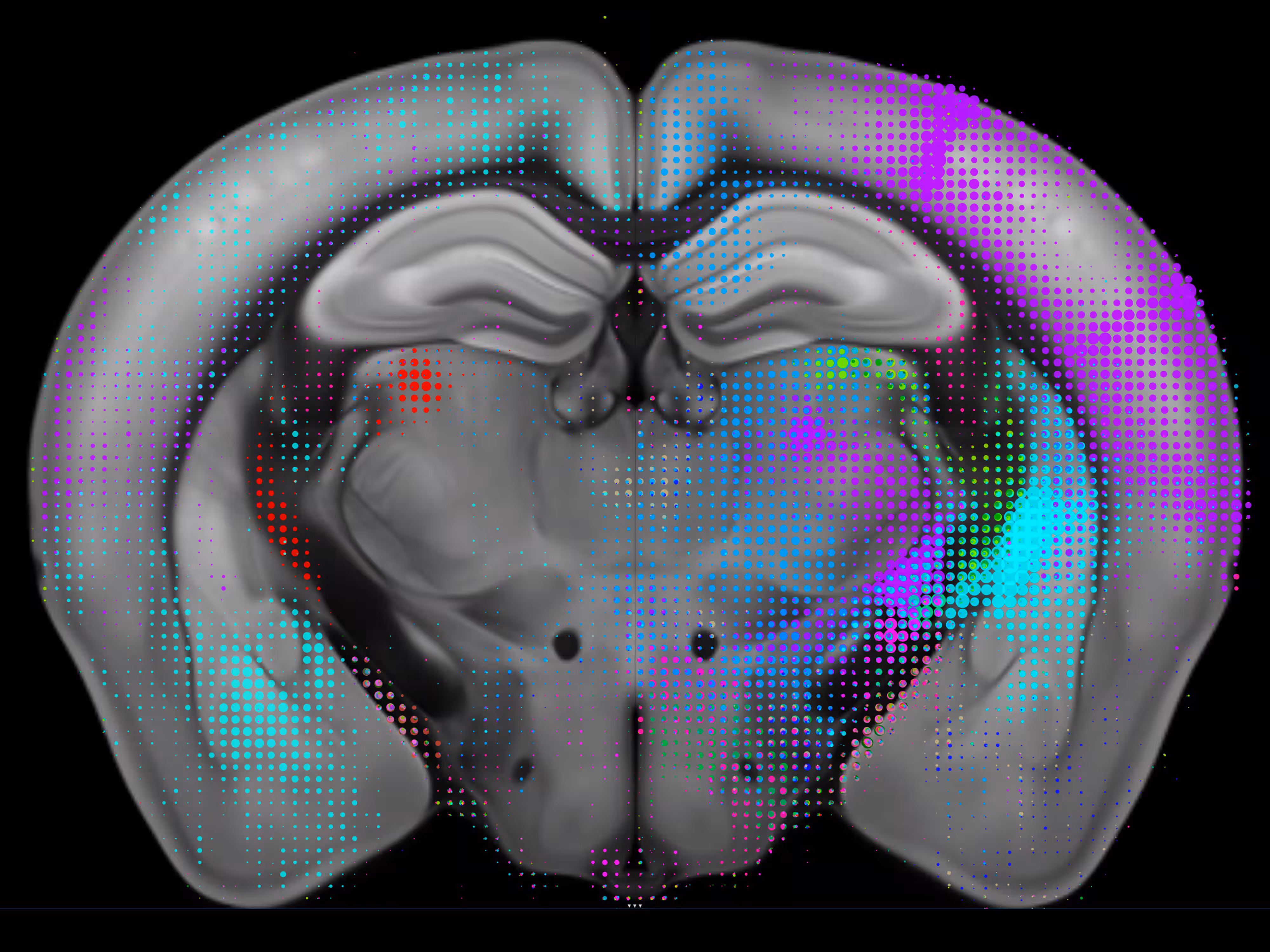
axonal connectomics

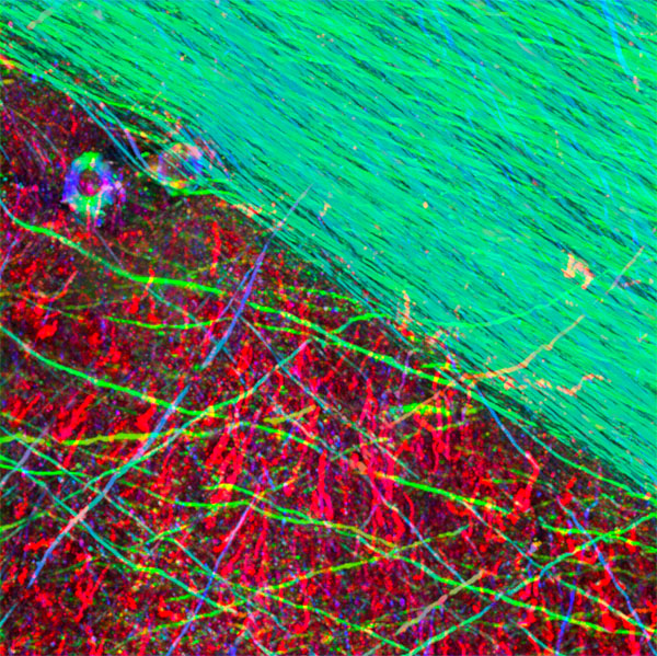
While the EM connectomics has formed the bulk of our research over the past decade, the newer Axonal Connectomics project aims to apply connectomics approaches to larger brains, with the complete human brain as the ultimate goal. While the EM approach can reasonably be applied to the entire mouse brain in the coming year, the required data sizes make it impossible to do EM imaging of entire human brain. With lower resolution, however, it is an achievable goal to map the majority of large (myelinated) axons in the coming years. Given that never has a single axon been traced from source to target through the human white matter, a new approach is clearly needed.

With a combination of antibody staining, tissue clearing, expansion, and light-sheet microscopy, we have begun mapping the courses of projection axons in large brains at high resolution. The resultant three-dimensional data sets are readily segmented both by humans and with machine-learning approaches, similar to those used with electron microscopy. We are currently working to scale-up our centimeter-scale data sets to increasingly large portions of larger brains, including the human.

‍

Vibrant abstract digital art with red, green, and neon colors forming intersecting patterns
Project Lead: Clay Reid

‍Team members: Soumya Chatterjee, Steven Cook, Ayana Hellevik, Cheryl Lea, Kevin Takasaki, Russel Torres, Emily Turschak, Kareena Villalobos, Wan-Qing Yu
‍
‍Non-departmental members: Olga Gliko, Uygar Sumbul

featured publications

explore more publications
publication / 2025
Inhibitory specificity from a connectomic census of mouse visual cortex
Nature
publication / 2025
Functional connectomics reveals general wiring rule in mouse visual cortex
Nature
publication / 2022
Reconstruction of neocortex: Organelles, compartments, cells, circuits, and activity
Cell
explore more publications

related news

all news
video
Revealing the largest wiring diagram and functional map of the brain through MICrONS
Scientists have created the largest wiring diagram and functional map of an animal brain
news
Attempting the impossible: A 20-year journey to learn the language of the brain
In what is considered the most complicated neuroscience experiment ever attempted, scientists from the Allen Institute and global collaborators have...
news
Scientists recreated part of the mouse brain on a computer - and showed it movies
The 230,000-cell simulation of part of the visual system can serve as a testbed for understanding general principles of the brain, its creators say

projects and data

Colorful neon neural network or interconnected nodes glowing against black background
The MICrONS Project
nature features an unprecedented dataset of high resolution anatomical images of individual cells in mouse visual cortex, mapped on to their responses
3D rendered neuron cell body with colorful branching dendrites and axons
MICrONS Dataset
open access data and visual resources to explore the dataset
3D mouse brain visualization with colorful pixelated overlay showing neural activity regions
Allen
Brain-map data access
access connectomics data on brain-map.org
CAVEconnectome logo with circular icon on dark background
Connectome Annotation Versioning Engine (CAVE)
CAVE is a computational infrastructure for immediate and reproducible connectome analysis in up-to petascale datasets (~ 1 m m 3 ) while proofreading and annotating is ongoing

connectomics team

Agnes L. Bodor
Scientist III
Derrick Brittain
SW Engineer III
Soumya Chatterjee
Scientist III
Forrest Collman
Associate Director, Data and Technology
Nuno Maçarico da Costa
Investigator
Bethanny Danskin
Scientist I
Kim Gruver
Scientist I
Cheryl Lea
Manager, Research Administration
Melissa Lerch
Scientific Project and Alliance Manager
Xiaoyu Lu
Scientist I
Ben Pedigo
Scientist I
R. Clay Reid
Investigator, Sr. Connectomics
Jenna Schardt
Research Associate, Sr.
Kevin Takasaki
Scientist III
Marc Takeno
Research Associate Principal
Russel Torres
SW Engineer III
Emily Turschak
Research Associate III (PT)
Kareena Villalobos
Research Associate 1
Keith Wiley
Connectomics Service Engineer
Wan-Qing Yu
Scientist II
Chi Zhang
Scientist II
we acceleratedevelopcatalyzeimpact

science done differently. shared openly.

explore our accelerators

brain science

Mapping every cell, connection, and circuit in the brain—openly shared with the world.

cell science

Decoding how cells become tissues, then programming that knowledge into powerful new research tools.

neural dynamics

Revealing the brain's hidden algorithms that transform neural activity into real-world behavior.

immunology

Creating the deepest open reference for the healthy human immune system ever built.

synthetic biology

Engineering cells to record their own histories, transforming how we understand disease over time.

research

Big questions, open answers, and science built to be shared.

education

Inspiring the next generation of scientists through open science resources.

impact

Our science is empowering researchers and advancing health worldwide.
advancing science through open, collaborative research
Get the allen institute newsletter
Stay informed on the latest breakthroughs in neuroscience, bioscience, and AI-driven research.
allen institute
impactpeople & teamscareers & opportunitiesalumnihistory & founder
science resources
allencell.orgallenimmunology.orgallenneuraldynamics.orgbrain-bican.orgbrain-map.orgmicrons-explorer.org
research
brain sciencecell scienceneural dynamicsimmunologysynthetic biologypublications
education
science educationfield tripsprofessional developmenteducation resources
quick links
newseventsopen sciencepodcastscience resourceshuman brain donationvisit uscontact
follow us/

allen institute, 615 Westlake Ave North, Seattle, WA 98109 +12065487055

© 0000 allen institute. all rights reserved.
privacy policyterms of usecitation policyemployee portalpolicy & compliancecookie settings