in this article
What are the parts of the brain?
Understanding the human mind
By Jenny Burns
Spinning 3D human brain from the Allen Human Brain Atlas
Your brain is amazing and complex
An intricate powerhouse that keeps you thinking, feeling, and functioning every day
Did you know that part of your brain is built from the inside out? Have you ever considered why the brain has a wrinkly exterior?
You may be familiar with terms like white matter, grey matter, neurons, and the cerebral cortex—and maybe you’ve wondered what they mean, where they are, or what they do.
Enjoy this visual and informative journey as we explore the brain’s many regions and parts. Scientists at the Allen Institute and around the world are working to uncover the secrets of this remarkable organ — the control center of the body and often considered the most complex object in the known universe.
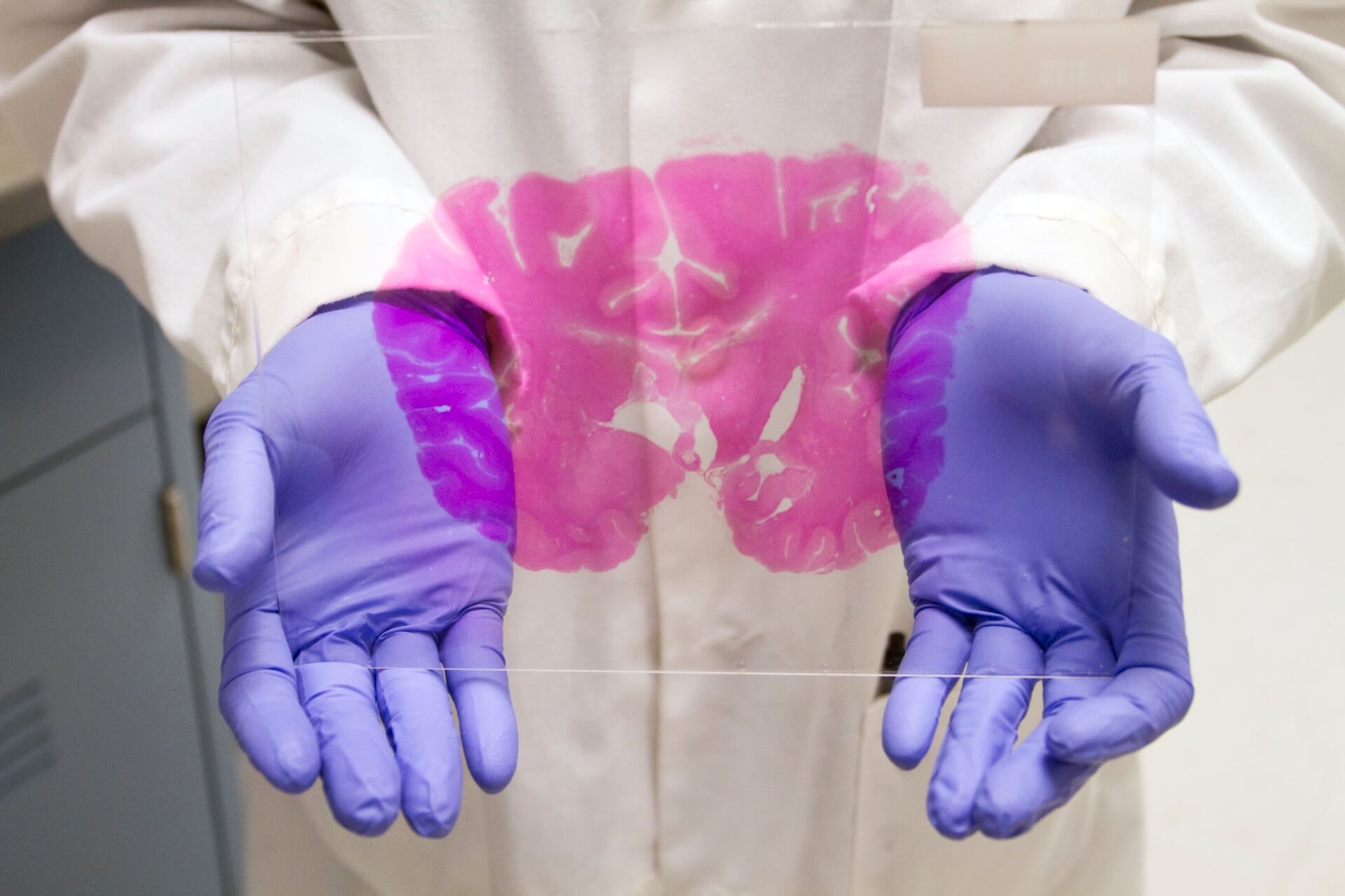
This image shows an Allen Institute scientist holding a section of human brain tissue used for research.
Understanding brain basics
Brain anatomy is divided into three main sections: the cerebrum, cerebellum, and brainstem. These parts work together to power everything from basic survival functions to complex thoughts. All vertebrates have these parts, but they differ widely in relative shape, size, and complexity. In the bird brainstem, there is a large area called the “optic tectum;” this area is important for processing visual information and spatial attention. Meanwhile, humans and other primates have very large cerebrums.
Within the brain, you’ll also find white matter and grey matter. White matter gets its name from the fatty myelin coating around axons, which helps signals travel between neurons faster—think of it as the brain’s communication highway. Grey matter, on the other hand, includes neuron cell bodies and dendrites, which are crucial for processing and integrating information.
The video shows stained human tissue (left) and highlighted anatomical structures (right) from the Allen Human Brain Atlas.
The Cerebrum
The cerebrum is the largest part of the human brain. It includes structures like the cerebral cortex, the outer-most portion of the cerebrum, that is responsible for things like sensation, motor planning, thoughts, and personality.
Fun fact: “cortex” is Latin for “bark” or “shell.”
The folds, or wrinkles, in the cerebral cortex serve an important function to increase the amount of brain surface area. Unlike birds and reptiles, most mammals have brain folding, with a few exceptions (e.g., mice).
Underneath the cerebral cortex, and still within the cerebrum, we have structures like the hippocampus — which is responsible for spatial navigation and memory, and the basal ganglia — which are responsible for reward, motivation, and movement. The basal ganglia are especially important for understanding Parkinson’s Disease (PD), a neurodegenerative disorder with movement difficulties, as this brain area contains unhealthy dopamine neurons in PD.
Cerebellum
The “mini brain”
The cerebellum, lovingly referred to as the “mini brain,” is responsible for a lot of things like balance, movement, and muscle memory. Maybe you’ve heard about “wobbly cat syndrome,” a condition where cats have an abnormally small cerebellum and poor balance. The cerebellum is located in the back of the brain, below the cerebrum and just above where your spinal cord connects to your brain.
Fun fact: The cerebellum has one of the largest cell types in the brain, called Purkinje neurons.
This Nissl image of a section of adult human brain tissue from the Allen Human Brain Atlas, shows structures of the cerebellum, located below the cerebrum in the back of the brain.
Brain Stem
The brain stem keeps you alive. It is responsible for a lot of things that you don’t even realize that you’re doing – breathing, swallowing, regulating your heart rate. Cranial nerves extend from the brainstem to your head, neck, and torso and contain sensory andmotor information. For example, the vagus nerve (cranial nerve 10), connects the brainstem to the heart, lungs, and digestive tract and is part of the “rest and digest” parasympathetic nervous system – this is why deep breathing calms the brain down.
The brain stem imagery is courtesy of Society for Neuroscience via brainfacts.org.
Stuff the brain is made of
A closer look at brain tissue
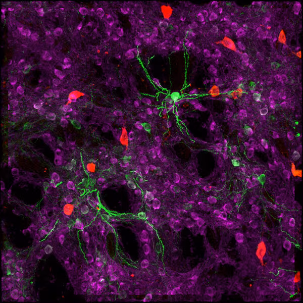
The brain is mostly made of fat, fluids, and cells. While scientists are interested in all of this matter, many of them have a particular interest in brain cells as they explore how cells impact learning, memory, healthy brain function, and disease. Brain cells are largely divided into neurons, the cells that send electrical signals, and non-neuronal support cells.
Brain cells can be further characterized into different types based on their electrical properties, their shape, the genes they express, and their connections to other cells. Chandelier cells and rosehip neurons are both examples of brain cell types that are named for their shape. Scientists at Allen Institute, together with global collaborators and supported by NIH funding, are creating a massive atlas of cell types in the human brain.
Fun fact: Neurons are born in the inner part of the brain and often migrate outwards past other neurons to get to their final location as the brain grows during development.
Neuroscience at Allen Institute
Unraveling the mysteries of the brain
The Allen Institute is an independent nonprofit bioscience research institute in Seattle, Washington dedicated to understanding life and advancing health.
We are world leaders in neuroscience, cell biology and immunology research. Our brain science teams are focused on examining brain cell types and their connections in healthy and diseased states – including Alzheimer’s disease, understanding how the brain’s activity leads to behavior, and examining consciousness in the mammalian brain.
Open science is a founding core research principle at the Allen Institute. We share our findings, data, educational resources, and tools openly to accelerate progress and drive breakthroughs worldwide.
Have you ever wondered: What is the brain? Where does it begin and end? What’s it made of, and how does it differ across species?
Sign up for our newsletter to keep up to date with news from the Allen Institute
Authors


Citations
about the allen institute
Allen Institute is a 501(c)(3) nonprofit medical research organization dedicated to accelerating science for a healthier world. Through large-scale, multidisciplinary research initiatives, the Institute generates foundational knowledge, data, tools, and models that are shared openly with the world to advance our understanding of life and health. Founded by Jody Allen and the late Paul G. Allen, Allen Institute is supported primarily by the Fund for Science and Technology.