ALS is a cruel disease. It robs the body of its ability to control itself—the ability to move, the ability to communicate. While there are currently no effective treatments to reverse its debilitating symptoms, Allen Institute researchers have opened a window of hope.
For the first time ever, scientists have developed a precise genetic toolkit that can target the exact nerve cells destroyed by the disease and potentially deliver therapies where they are needed most—a discovery that could dramatically speed up the quest for a cure. The findings were recently published in the journal Cell Reports.
Why it Matters
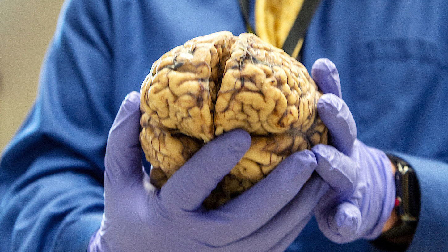
Amyotrophic lateral sclerosis (ALS) is a progressive and devastating disease that gradually kills off motor neurons in the brain and spinal cord that control voluntary muscle movement. As these neurons die, people with ALS lose the ability to move, speak, and eventually breathe. Despite decades of research, there’s still no effective treatment or cure. Unlike many other brain cells, motor neurons in the spinal cord have been extremely hard to reach with genetic tools. This has slowed down research and made it hard to test new treatments in the cells that matter most.
This new genetic tool acts like a “GPS system” to specifically find and label motor neurons—without affecting any other type of cell—offering a new way to study ALS from the inside out and deliver therapeutic agents that could prevent the cells connected to the disease from dying.
How Scientists Did It
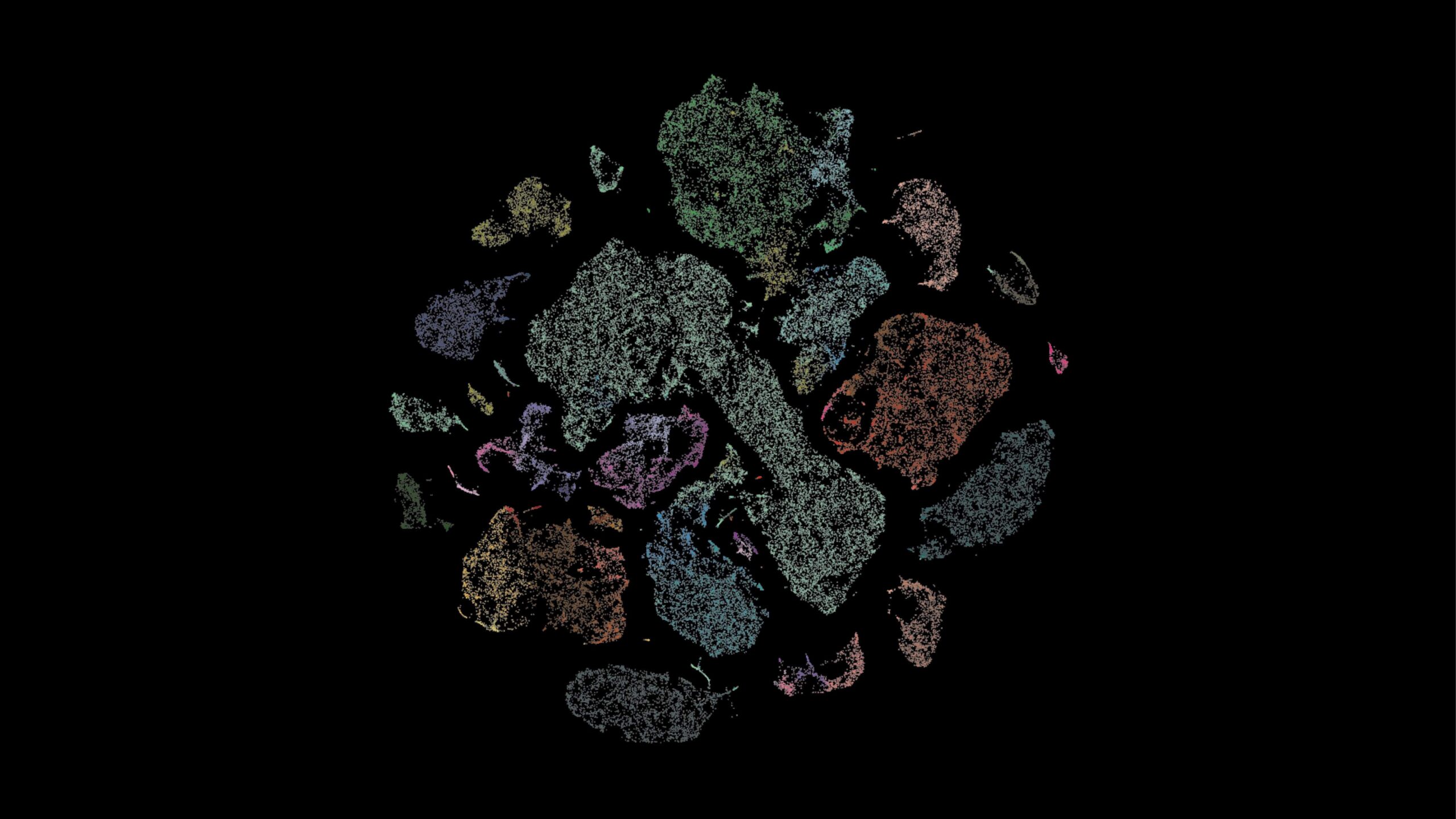
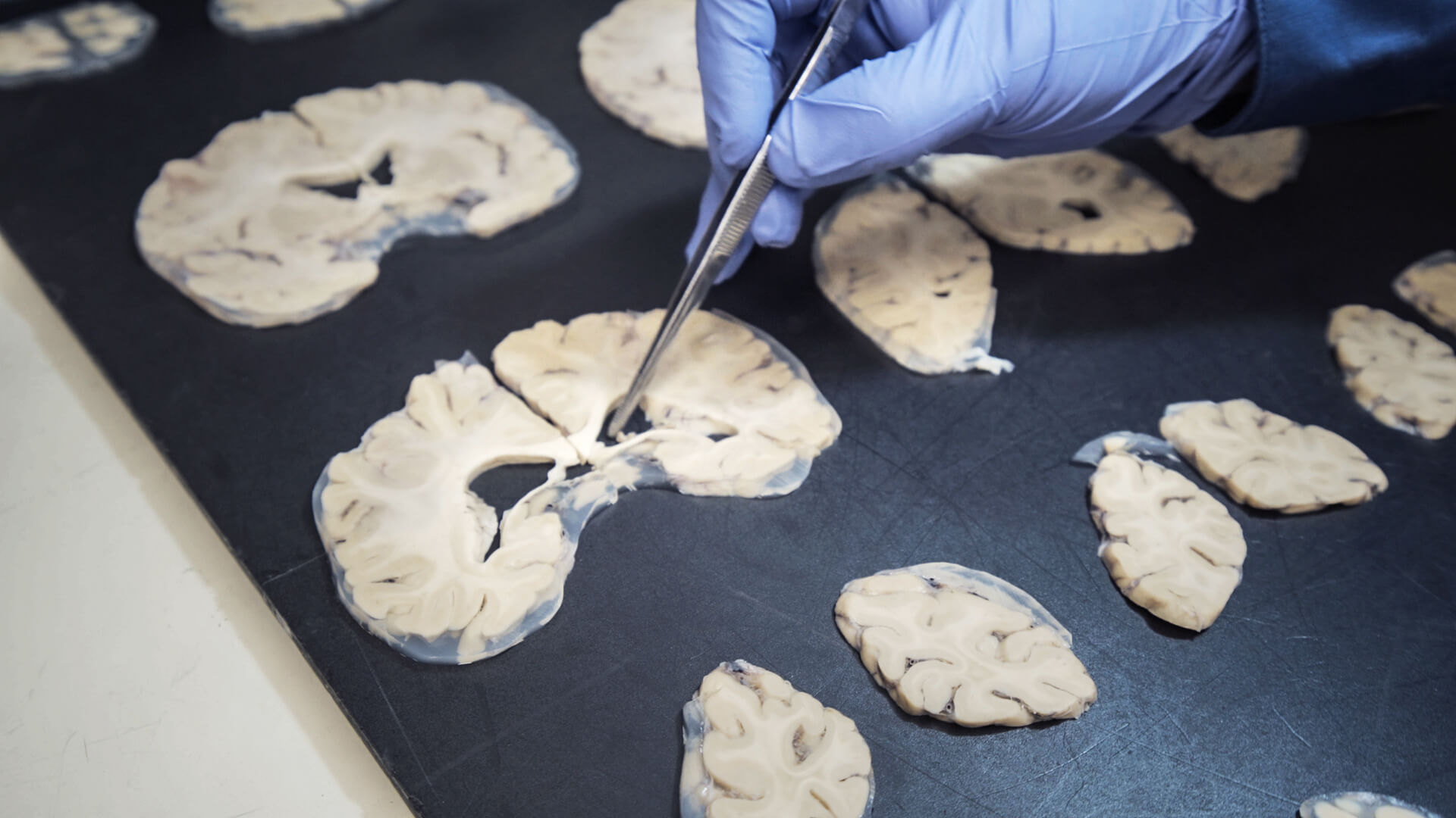
Enhancer AAVs are one type of genetic tool. Scientists began by creating a cellular map by analyzing spinal cord tissue from different species, looking at each cell individually and analyzing their molecular makeup. From that map they found short pieces of DNA, known as enhancers, that work like on/off switches in cells, changing their gene expression and in turn their function or properties in some way.
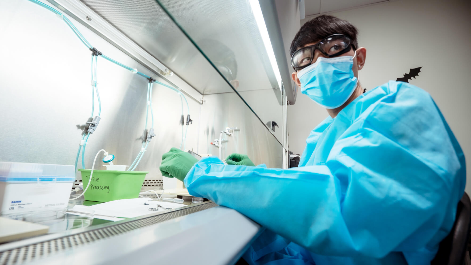
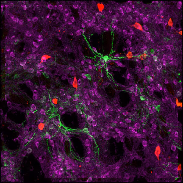
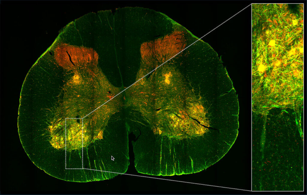
These enhancers activate only in specific neurons, making them the perfect tools for delivering targeted treatments or labels to those cells. Researchers then inserted these genetic switches into harmless viruses, along with a glowing protein that lights up under special microscopes to label the cells and verify that the targeting was successfully. These harmless viruses simply act as “delivery trucks” to send a therapeutic or functional cargo into the cell.
What this Could Mean for People with ALS
In the short term, researchers can now study how ALS develops in animal models, offering a better understanding of how the disease works. This could lead to new drug targets or biomarkers that tell doctors how fast the disease is progressing.
Eventually, this same system could be used to deliver gene therapies directly to motor neurons in the brain to protect them and prevent them from dying. Because it targets only the affected neurons, potential treatments could come with fewer side effects.
The toolkit could even pave the way for personalized ALS treatments. By studying different types of motor neurons, researchers might learn why ALS progresses differently in different people, and then tailor therapies to match. Right now, researchers are looking for partnerships to take these tools to the clinical space and fast track them for trials to start in just a few years.
“I know that timeline does sound long, and patients need something right now,” said Tanya Daigle, Ph.D., lead author on the research and associate investigator at the Allen Institute. “But the average time in drug development is about 15 years, so this would be astonishing progress.”
A Hopeful Sign for Success in Humans
What makes the findings exciting is that the genetic tools worked across multiple animal species, which increases the likelihood they will also work in humans. The “delivery trucks” successfully found and labeled motor neurons, lighting them up, while leaving other cells untouched. Scientists were also able to combine multiple enhancers to build a single tool to target two types of motor neurons: those in the brain and those in the spinal cord. Both fall victim to ALS, so reaching them at once is a major scientific advance.
This discovery gives researchers a new way forward. For decades, motor neurons have been largely out of reach, leaving scientists with few research options. With this new genetic toolkit, they have a better roadmap to find answers—and possibly even cures. For people living with ALS and their loved ones, it brings something that’s been in short supply: hope.
“I think it’s an exciting time to be a molecular neuroscientist,” said Daigle. “I became a scientist for exactly this moment in time, when something we do in the lab could benefit the people dealing with this horrible disease. It robs people of their loved ones—their husbands, fathers, wives, mothers. And I think that’s why a lot of us in science become scientists. The potential to impact humanity like this is a very exciting thing.”
Citations
about the allen institute
Allen Institute is a 501(c)(3) nonprofit medical research organization dedicated to accelerating science for a healthier world. Through large-scale, multidisciplinary research initiatives, the Institute generates foundational knowledge, data, tools, and models that are shared openly with the world to advance our understanding of life and health. Founded by Jody Allen and the late Paul G. Allen, Allen Institute is supported primarily by the Fund for Science and Technology.