.avif)

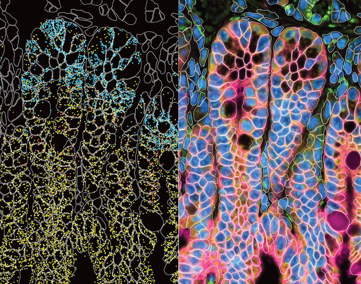
This vibrant, rainbow-like mosaic of colors arranged in irregular patterns shows millions of cells in the human body and how they're related to one another. Each tiny dot represents a single cell; and their color, the cell type: cyan for intestinal cells, red for T cells, white for liver cells, orange for smooth muscle cells.
The shapes and patterns they form reveal how similar or different each cell is to another based off the genes that are turned on inside of them (gene expression). When a gene is turned on, or active, it’s performing a specific function in the body. The closer one dot is to another, the more similar is the gene expression of the cells they represent.
The technical name for this vibrant image is a Uniform Manifold Approximation and Projection (UMAP). Researchers use UMAPs to simplify and visualize complex data so that it’s more understandable and interpretable.
In the video, the UMAP transitions into 15 distinct spatial maps. Each dot (or cell) reforms into a cross-section of tissue, revealing their exact location within that organ. For example, the top middle image represents a cross-section of the intestine. The cyan color represents the absorptive enterocytes (intestinal cells) forming the mucosal lining, while the orange identifies the underlying smooth muscle layer.

“What we’re trying to combine is the structural information of microscopy with the transcriptomic information of single-cell sequencing,” said Maximilian Heeg, assistant investigator at the Allen Institute. “It allows us to see how they interact with other cells to make meaning of these bigger ecosystems instead of just looking at one cell.”
These visualizations are an example of spatial transcriptomics.
what is spatial transcriptomics?
Spatial transcriptomics is a technique that measures what genes are turned on, or active, in a cell and where that cell is located within a sample of tissue. Instead of just knowing what cells are there, it lets researchers see how those cells are organized and how they talk to their neighbors. It’s vital for immunologists
“With this technology, we can put the immune system in tissues into a new context, which we hope will change how we think about regulating or targeting immune responses in tissues,” said Heeg.
Scientists can learn certain things about a cell in isolation. But like a single word taken out of context compared with its usage in a sentence, cell characteristics take on more meaning when they’re surrounded by other cells and put into spatial context (i.e. their physical location) in relation to other cells.
Citations
about the allen institute
The Allen Institute is an independent, 501(c)(3) nonprofit research organization founded by philanthropist and visionary, the late Paul G. Allen. The Allen Institute is dedicated to answering some of the biggest questions in bioscience and accelerating research worldwide. The Institute is a recognized leader in large-scale research with a commitment to an open science model. For more information, visit alleninstitute.org.